This old version of Proteopedia is provided for student assignments while the new version is undergoing repairs. Content and edits done in this old version of Proteopedia after March 1, 2026 will eventually be lost when it is retired in about June of 2026.
Apply for new accounts at the new Proteopedia. Your logins will work in both the old and new versions.
NAD synthase
From Proteopedia
(Difference between revisions)
| (7 intermediate revisions not shown.) | |||
| Line 1: | Line 1: | ||
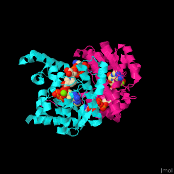
| - | <StructureSection load=' | + | <StructureSection load='' size='350' side='right' caption='NAD+ synthase complex with Mg+2 (green), ATP and deamino-NAD (PDB entry [[1xng]])' scene='54/541072/Cv/1'> |
== Function == | == Function == | ||
| - | '''NAD+ synthase''' (NADS) catalyzes the conversion of ATP, deamino-NAD+ and NH<sub>3</sub> to AMP, NAD+ and diphosphate (PP). NADS is part of the NAD synthesis pathway<ref>PMID:12898714</ref> . See [[NAD]]. | + | '''NAD+ synthase''' or '''NH3-dependent NAD synthetase''' (NADS) catalyzes the conversion of ATP, deamino-NAD+ and NH<sub>3</sub> to AMP, NAD+ and diphosphate (PP). NADS is part of the NAD synthesis pathway<ref>PMID:12898714</ref> . See [[NAD]]. |
== Structural highlights == | == Structural highlights == | ||
| - | The <scene name='54/541072/Cv/ | + | The <scene name='54/541072/Cv/8'>ATP binding site</scene> contains <scene name='54/541072/Cv/9'>Mg+2 ion</scene>. The <scene name='54/541072/Cv/10'>NAD binding site is located in the interface of the 2 subunits</scene><ref>PMID:15645437</ref>. Water molecules are shown as red spheres. |
</StructureSection> | </StructureSection> | ||
== 3D Structures of NAD+ synthase == | == 3D Structures of NAD+ synthase == | ||
| Line 20: | Line 20: | ||
**[[2pzb]] – BaNADS - ''Bacillus anthracis'' <br /> | **[[2pzb]] – BaNADS - ''Bacillus anthracis'' <br /> | ||
**[[3dpi]] – NADS – ''Burkholderia pseudomallei''<br /> | **[[3dpi]] – NADS – ''Burkholderia pseudomallei''<br /> | ||
| + | **[[6kv3]] – NADS – ''Staphylococcus aureus''<br /> | ||
| + | **[[5wp0]] – NADS – ''Allivibrio fischeri''<br /> | ||
| + | **[[5huh]], [[5huj]] – NADS – ''Streptococcus pyogenes''<br /> | ||
| + | **[[3n05]] – NADS – ''Streptomyces avermitillis''<br /> | ||
| + | **[[4xfd]] – PaNADS – ''Pseudomonas aeruginosa''<br /> | ||
| + | **[[4q16]] – NADS – ''Deinococcus radiodurans''<br /> | ||
| + | **[[2e18]] – NADS – ''Pyrococcus horikoshii''<br /> | ||
*NADS binary complex | *NADS binary complex | ||
| Line 26: | Line 33: | ||
**[[1wxh]] – EcNADS + NAD <br /> | **[[1wxh]] – EcNADS + NAD <br /> | ||
**[[3hmq]] – BsNADS + NAD – ''Salmonella typhimurium'' <br /> | **[[3hmq]] – BsNADS + NAD – ''Salmonella typhimurium'' <br /> | ||
| + | **[[6c8q]] – NADS + NAD – ''Enterococcus faecalis''<br /> | ||
| + | **[[5f23]] – PaNADS + NAD <br /> | ||
*NADS ternary complex | *NADS ternary complex | ||
| Line 33: | Line 42: | ||
**[[1wxg]] – EcNADS + Mg + deamino-NAD <br /> | **[[1wxg]] – EcNADS + Mg + deamino-NAD <br /> | ||
**[[2pz8]] – BaNADS + Mg + ATP derivative <br /> | **[[2pz8]] – BaNADS + Mg + ATP derivative <br /> | ||
| + | **[[3fiu]] – NADS + Mg + AMP – ''Francisella tularensis''<br /> | ||
*NADS quaternary complex | *NADS quaternary complex | ||
| Line 50: | Line 60: | ||
*NADS glutamine-dependent | *NADS glutamine-dependent | ||
| + | **[[6ofb]] – hNADS + AMP + NAD derivative<br /> | ||
**[[3sdb]] – MtNADS (mutant) – ''Mycobacterium tuberculosis'' <br /> | **[[3sdb]] – MtNADS (mutant) – ''Mycobacterium tuberculosis'' <br /> | ||
**[[3seq]], [[3sez]] – MtNADS (mutant) + ATP derivative + pyridinium derivative <br /> | **[[3seq]], [[3sez]] – MtNADS (mutant) + ATP derivative + pyridinium derivative <br /> | ||
**[[3szg]] – MtNADS (mutant) + AMP + pyridinium derivative <br /> | **[[3szg]] – MtNADS (mutant) + AMP + pyridinium derivative <br /> | ||
**[[3syt]] – MtNADS + AMP + NAD + glutamate <br /> | **[[3syt]] – MtNADS + AMP + NAD + glutamate <br /> | ||
| + | **[[3dla]] – MtNADS + norleucine + deamido-NAD <br /> | ||
| + | **[[6ofc]] – MtNADS (mutant) + AMP + sulfonamide derivative + glutamate <br /> | ||
| + | **[[4f4h]] – NADS – ''Burkholderia thailandensis''<br /> | ||
| + | **[[3ilv]] – NADS – ''Cytophaga hutchinsonii''<br /> | ||
}} | }} | ||
Current revision
| |||||||||||
3D Structures of NAD+ synthase
Updated on 15-November-2021
References
- ↑ Suda Y, Tachikawa H, Yokota A, Nakanishi H, Yamashita N, Miura Y, Takahashi N. Saccharomyces cerevisiae QNS1 codes for NAD(+) synthetase that is functionally conserved in mammals. Yeast. 2003 Aug;20(11):995-1005. PMID:12898714 doi:http://dx.doi.org/10.1002/yea.1008
- ↑ Kang GB, Kim YS, Im YJ, Rho SH, Lee JH, Eom SH. Crystal structure of NH3-dependent NAD+ synthetase from Helicobacter pylori. Proteins. 2005 Mar 1;58(4):985-8. PMID:15645437 doi:10.1002/prot.20377
