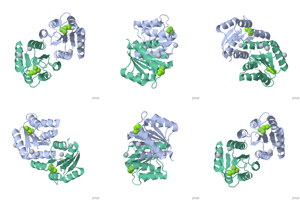
PhoP-PhoQ
From Proteopedia
(Difference between revisions)
| (3 intermediate revisions not shown.) | |||
| Line 12: | Line 12: | ||
---- | ---- | ||
| - | E. coli phoP is a 223 residue protein containing a 120-residue regulatory domain joined by a 5-residue linker to a 98 residue c-terminal DNA binding effector domain.<ref name=Bachhawat>PMID:17545283</ref> At a conserved Asp residue (<scene name='PhoP-PhoQ/Asp51-bef3/1'>Asp51 in this case</scene>), the regulatory domain can be modified by a phosphoryl group from the protein kinase function of phoQ. Phosphorylation of the regulatory domain modulates the activity of the effector domain to bind DNA and regulate transcription. Phosphorylated, or "activated" phoP binds to "phoP boxes" on bacterial DNA, which consist of two direct hexanucleotide repeats separated by a five nucleotide spacer located at the -35 position.<ref name=Bachhawat>PMID:17545283</ref>: | + | ''E. coli'' phoP is a 223 residue protein containing a 120-residue regulatory domain joined by a 5-residue linker to a 98 residue c-terminal DNA binding effector domain.<ref name=Bachhawat>PMID:17545283</ref> At a conserved Asp residue (<scene name='PhoP-PhoQ/Asp51-bef3/1'>Asp51 in this case</scene>), the regulatory domain can be modified by a phosphoryl group from the protein kinase function of phoQ. Phosphorylation of the regulatory domain modulates the activity of the effector domain to bind DNA and regulate transcription. Phosphorylated, or "activated" phoP binds to "phoP boxes" on bacterial DNA, which consist of two direct hexanucleotide repeats separated by a five nucleotide spacer located at the -35 position.<ref name=Bachhawat>PMID:17545283</ref>: |
(T/G)GTTTA | (T/G)GTTTA | ||
| Line 32: | Line 32: | ||
Spontaneous dimerization of the unactivated phoP regulatory domain can be observed in vitro, but is likely due to high concentration of the protein and may not occur in vivo. Although the dimerization of the unactivated phoP regulatory domain results in a homodimer similar to the activated homodimer, it is significantly less stable. Activation by the phosphoryl group helps to stabilize the <scene name='Sandbox_Reserved_344/Dimerization_surface/1'>α-4 helix, β-5 sheet and α-5 helix face</scene> dimerization interphase. Without the phosphoryl group, the two monomers dimerize in an asymmetric fashion that lends to molecular instability. | Spontaneous dimerization of the unactivated phoP regulatory domain can be observed in vitro, but is likely due to high concentration of the protein and may not occur in vivo. Although the dimerization of the unactivated phoP regulatory domain results in a homodimer similar to the activated homodimer, it is significantly less stable. Activation by the phosphoryl group helps to stabilize the <scene name='Sandbox_Reserved_344/Dimerization_surface/1'>α-4 helix, β-5 sheet and α-5 helix face</scene> dimerization interphase. Without the phosphoryl group, the two monomers dimerize in an asymmetric fashion that lends to molecular instability. | ||
| - | + | ||
===PhoP-PhoQ and Virulence=== | ===PhoP-PhoQ and Virulence=== | ||
| Line 44: | Line 44: | ||
Two component regulatory systems such as phoP-phoQ are obviously an attractive target for future antimicrobial drugs. If the phoP-phoQ can be altered to a dysfunctional state, the relevant bacteria would have decreased pathogenicity and increased suceptibility to nonspecific immune defense. | Two component regulatory systems such as phoP-phoQ are obviously an attractive target for future antimicrobial drugs. If the phoP-phoQ can be altered to a dysfunctional state, the relevant bacteria would have decreased pathogenicity and increased suceptibility to nonspecific immune defense. | ||
| + | </StructureSection> | ||
== 3D Structures of PhoP-PhoQ== | == 3D Structures of PhoP-PhoQ== | ||
Updated on {{REVISIONDAY2}}-{{MONTHNAME|{{REVISIONMONTH}}}}-{{REVISIONYEAR}} | Updated on {{REVISIONDAY2}}-{{MONTHNAME|{{REVISIONMONTH}}}}-{{REVISIONYEAR}} | ||
| Line 61: | Line 62: | ||
**[[3cgz]] - StPhoQ catalytic domain<br /> | **[[3cgz]] - StPhoQ catalytic domain<br /> | ||
**[[1yax]] - StPhoQ sensor domain (mutant)<br /> | **[[1yax]] - StPhoQ sensor domain (mutant)<br /> | ||
| - | **[[3bq8]] – EcPhoQ<br /> | + | **[[3bq8]], [[6a8u]] – EcPhoQ sensor domain<br /> |
| - | **[[3bqa]] – EcPhoQ (mutant)<br /> | + | **[[3bqa]], [[6a8v]] – EcPhoQ sensor domain (mutant)<br /> |
| - | **[[1id0]] – EcPhoQ kinase domain | + | **[[1id0]] – EcPhoQ kinase domain<br /> |
| + | **[[4uey]] - PhoQ periplasmic domain (mutant) – ''Salmonella enterica''<br /> | ||
}} | }} | ||
===References=== | ===References=== | ||
Current revision
| |||||||||||
3D Structures of PhoP-PhoQ
Updated on 22-September-2020
References
- ↑ 1.0 1.1 1.2 Hoch JA. Two-component and phosphorelay signal transduction. Curr Opin Microbiol. 2000 Apr;3(2):165-70. PMID:10745001
- ↑ 2.0 2.1 2.2 2.3 2.4 Bachhawat P, Stock AM. Crystal structures of the receiver domain of the response regulator PhoP from Escherichia coli in the absence and presence of the phosphoryl analog beryllofluoride. J Bacteriol. 2007 Aug;189(16):5987-95. Epub 2007 Jun 1. PMID:17545283 doi:10.1128/JB.00049-07
- ↑ Angelichio MJ, Camilli A. In vivo expression technology. Infect Immun. 2002 Dec;70(12):6518-23. PMID:12438320

