We apologize for Proteopedia being slow to respond. For the past two years, a new implementation of Proteopedia has been being built. Soon, it will replace this 18-year old system. All existing content will be moved to the new system at a date that will be announced here.
Galactose-binding lectin
From Proteopedia
(Difference between revisions)
| (13 intermediate revisions not shown.) | |||
| Line 1: | Line 1: | ||
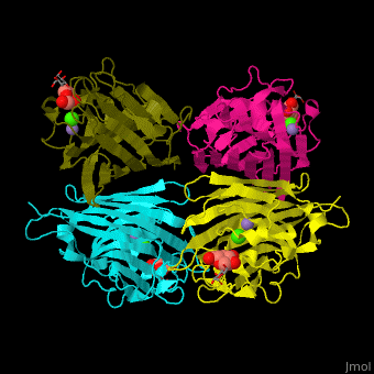
| - | <StructureSection load='' size=' | + | <StructureSection load='' size='450' side='right' caption='Structure of peanut galactose-binding lectin tetramer complex with β-D-galactose, α-D-glucose, Ca+2 (green) and Mn+2 (purple) ions (PDB entry [[1v6i]])' scene='57/572274/Cv/1'> |
| - | + | ||
== Function == | == Function == | ||
| Line 11: | Line 11: | ||
== Structural highlights == | == Structural highlights == | ||
| - | The carbohydrate ligand binds to 3 loops<ref>PMID:14747696</ref>. <scene name='57/572274/Cv/ | + | The carbohydrate ligand binds to 3 loops<ref>PMID:14747696</ref>. <scene name='57/572274/Cv/6'>β-D-galactose/α-D-glucose binding site</scene> in peanut galactose-binding lectin (PDB entry [[1v6i]]). |
| - | <scene name='57/572274/Cv/ | + | <scene name='57/572274/Cv/7'>Ca coordination site</scene>. |
| - | <scene name='57/572274/Cv/ | + | <scene name='57/572274/Cv/8'>Mn coordination site</scene>. Water molecules shown as red spheres. |
</StructureSection> | </StructureSection> | ||
| Line 32: | Line 32: | ||
**[[2dv9]], [[2dvd]] – GBL + β-D-galactose <br /> | **[[2dv9]], [[2dvd]] – GBL + β-D-galactose <br /> | ||
**[[1rir]] – GBL + porphyrin derivative <br /> | **[[1rir]] – GBL + porphyrin derivative <br /> | ||
| + | **[[6v95]], [[6vav]], [[6vaw]], [[6vc3]], [[6vc4]], [[6vgf]] – GBL + galactopyranose derivative <br /> | ||
*''Peanut GBL ternary complex'' | *''Peanut GBL ternary complex'' | ||
| Line 48: | Line 49: | ||
**[[1l7l]], [[1uoj]] – GBL <br /> | **[[1l7l]], [[1uoj]] – GBL <br /> | ||
| + | **[[4cp9]], [[4cpb]], [[7z62]], [[7z63]], [[8guv]], [[9g3r]] – GBL + galactoside <br /> | ||
| + | **[[7fio]], [[7fjh]] – GBL + inhibitor<br /> | ||
*''PaGBL binary complex'' | *''PaGBL binary complex'' | ||
| Line 53: | Line 56: | ||
**[[2wyf]] – GBL + methyl-β-galactose <br /> | **[[2wyf]] – GBL + methyl-β-galactose <br /> | ||
**[[4ljh]] – GBL + β-D-galactose <br /> | **[[4ljh]] – GBL + β-D-galactose <br /> | ||
| - | **[[3zyf]], [[4a6s]] – GBL + galactopyranose derivative<br /> | + | **[[3zyf]], [[4a6s]], [[4yw6]], [[5mih]] – GBL + galactopyranose derivative<br /> |
| + | **[[4ywa]] – GBL + triazole derivative <br /> | ||
| + | **[[6yo3]], [[6yoh]] – GBL + oxidanyl derivative <br /> | ||
*''PaGBL ternary complex'' | *''PaGBL ternary complex'' | ||
**[[3zyh]] – GBL + galactopyranose derivative + gal-lys-pro-leu <br /> | **[[3zyh]] – GBL + galactopyranose derivative + gal-lys-pro-leu <br /> | ||
| + | **[[4yw7]] – GBL + galactopyranose derivative + triazole derivative <br /> | ||
**[[3zyb]] – GBL + β-D-galactose + gal-lys-pro-leu<br /> | **[[3zyb]] – GBL + β-D-galactose + gal-lys-pro-leu<br /> | ||
**[[4lk6]] – GBL + β-D-galactose + chlorophenol red<br /> | **[[4lk6]] – GBL + β-D-galactose + chlorophenol red<br /> | ||
| Line 76: | Line 82: | ||
**[[2ds0]] – LtGBL C terminal (mutant) + sialyllactose <br /> | **[[2ds0]] – LtGBL C terminal (mutant) + sialyllactose <br /> | ||
**[[2zqo]] – LtGBL C terminal + GalNac <br /> | **[[2zqo]] – LtGBL C terminal + GalNac <br /> | ||
| + | |||
| + | *Other GBL | ||
| + | |||
| + | **[[5x4a]], [[3wmp]], [[3wmq]] – GBL residues 47-140 + polysaccharide – ''Sinularia lochmodes'' <br /> | ||
| + | **[[2zqn]] – eGBL C-terminal + lactose – earthworm<br /> | ||
| + | **[[2zqo]] – eGBL C-terminal + GalNac<br /> | ||
| + | **[[8vck]] – muGBL - mussell<br /> | ||
| + | **[[8vcm]], [[8vco]], [[8vcp]], [[8vcq]], [[8vcs]], [[8vcu]] – muGBL + galactoside<br /> | ||
| + | **[[6yf6]] – GBL C-terminal + fucoside derivative – ''Enterobacter cloacae''<br /> | ||
| + | |||
}} | }} | ||
== References == | == References == | ||
<references/> | <references/> | ||
[[Category:Topic Page]] | [[Category:Topic Page]] | ||
Current revision
| |||||||||||
3D structures of galactose-binding lectin
Updated on 10-July-2025
References
- ↑ Natchiar SK, Srinivas O, Mitra N, Surolia A, Jayaraman N, Vijayan M. Structural studies on peanut lectin complexed with disaccharides involving different linkages: further insights into the structure and interactions of the lectin. Acta Crystallogr D Biol Crystallogr. 2006 Nov;62(Pt 11):1413-21. Epub 2006, Oct 18. PMID:17057347 doi:10.1107/S0907444906035712
- ↑ Kundhavai Natchiar S, Arockia Jeyaprakash A, Ramya TN, Thomas CJ, Suguna K, Surolia A, Vijayan M. Structural plasticity of peanut lectin: an X-ray analysis involving variation in pH, ligand binding and crystal structure. Acta Crystallogr D Biol Crystallogr. 2004 Feb;60(Pt 2):211-9. Epub 2004, Jan 23. PMID:14747696 doi:http://dx.doi.org/10.1107/S090744490302849X
Proteopedia Page Contributors and Editors (what is this?)
Michal Harel, Alexander Berchansky, Joel L. Sussman, Jaime Prilusky
