|
|
| Line 1: |
Line 1: |
| | | | |
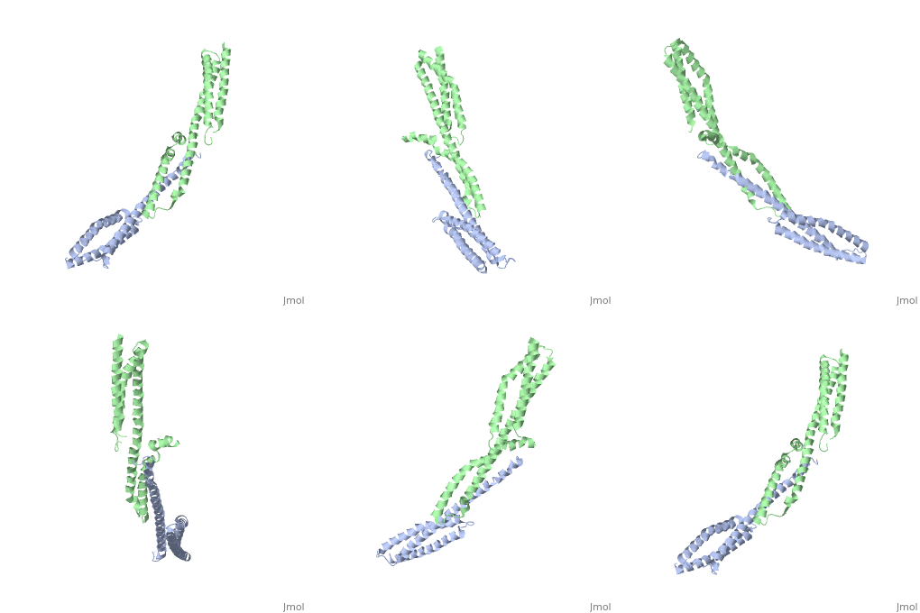
| | ==Crystal Structure of the Erythrocyte Spectrin Tetramerization Domain Complex== | | ==Crystal Structure of the Erythrocyte Spectrin Tetramerization Domain Complex== |
| - | <StructureSection load='3lbx' size='340' side='right' caption='[[3lbx]], [[Resolution|resolution]] 2.80Å' scene=''> | + | <StructureSection load='3lbx' size='340' side='right'caption='[[3lbx]], [[Resolution|resolution]] 2.80Å' scene=''> |
| | == Structural highlights == | | == Structural highlights == |
| - | <table><tr><td colspan='2'>[[3lbx]] is a 2 chain structure with sequence from [http://en.wikipedia.org/wiki/Human Human]. Full crystallographic information is available from [http://oca.weizmann.ac.il/oca-bin/ocashort?id=3LBX OCA]. For a <b>guided tour on the structure components</b> use [http://oca.weizmann.ac.il/oca-docs/fgij/fg.htm?mol=3LBX FirstGlance]. <br> | + | <table><tr><td colspan='2'>[[3lbx]] is a 2 chain structure with sequence from [https://en.wikipedia.org/wiki/Homo_sapiens Homo sapiens]. Full crystallographic information is available from [http://oca.weizmann.ac.il/oca-bin/ocashort?id=3LBX OCA]. For a <b>guided tour on the structure components</b> use [https://proteopedia.org/fgij/fg.htm?mol=3LBX FirstGlance]. <br> |
| - | </td></tr><tr id='related'><td class="sblockLbl"><b>[[Related_structure|Related:]]</b></td><td class="sblockDat">[[1owa|1owa]], [[3f31|3f31]], [[2spc|2spc]], [[1aj3|1aj3]], [[3edv|3edv]], [[3kbt|3kbt]]</td></tr> | + | </td></tr><tr id='method'><td class="sblockLbl"><b>[[Empirical_models|Method:]]</b></td><td class="sblockDat" id="methodDat">X-ray diffraction, [[Resolution|Resolution]] 2.8Å</td></tr> |
| - | <tr id='gene'><td class="sblockLbl"><b>[[Gene|Gene:]]</b></td><td class="sblockDat">SPTA, SPTA1 ([http://www.ncbi.nlm.nih.gov/Taxonomy/Browser/wwwtax.cgi?mode=Info&srchmode=5&id=9606 HUMAN]), SPTB, SPTB1 ([http://www.ncbi.nlm.nih.gov/Taxonomy/Browser/wwwtax.cgi?mode=Info&srchmode=5&id=9606 HUMAN])</td></tr>
| + | <tr id='resources'><td class="sblockLbl"><b>Resources:</b></td><td class="sblockDat"><span class='plainlinks'>[https://proteopedia.org/fgij/fg.htm?mol=3lbx FirstGlance], [http://oca.weizmann.ac.il/oca-bin/ocaids?id=3lbx OCA], [https://pdbe.org/3lbx PDBe], [https://www.rcsb.org/pdb/explore.do?structureId=3lbx RCSB], [https://www.ebi.ac.uk/pdbsum/3lbx PDBsum], [https://prosat.h-its.org/prosat/prosatexe?pdbcode=3lbx ProSAT]</span></td></tr> |
| - | <tr id='resources'><td class="sblockLbl"><b>Resources:</b></td><td class="sblockDat"><span class='plainlinks'>[http://oca.weizmann.ac.il/oca-docs/fgij/fg.htm?mol=3lbx FirstGlance], [http://oca.weizmann.ac.il/oca-bin/ocaids?id=3lbx OCA], [http://pdbe.org/3lbx PDBe], [http://www.rcsb.org/pdb/explore.do?structureId=3lbx RCSB], [http://www.ebi.ac.uk/pdbsum/3lbx PDBsum], [http://prosat.h-its.org/prosat/prosatexe?pdbcode=3lbx ProSAT]</span></td></tr> | + | |
| | </table> | | </table> |
| | == Disease == | | == Disease == |
| - | [[http://www.uniprot.org/uniprot/SPTA1_HUMAN SPTA1_HUMAN]] Defects in SPTA1 are the cause of elliptocytosis type 2 (EL2) [MIM:[http://omim.org/entry/130600 130600]]. EL2 is a Rhesus-unlinked form of hereditary elliptocytosis, a genetically heterogeneous, autosomal dominant hematologic disorder. It is characterized by variable hemolytic anemia and elliptical or oval red cell shape.<ref>PMID:2794061</ref> <ref>PMID:8018926</ref> <ref>PMID:1679439</ref> <ref>PMID:1878597</ref> <ref>PMID:2568862</ref> <ref>PMID:1541680</ref> <ref>PMID:8364215</ref> <ref>PMID:2384601</ref> <ref>PMID:1638030</ref> <ref>PMID:2568861</ref> <ref>PMID:8193371</ref> <ref>PMID:7772539</ref> Defects in SPTA1 are a cause of hereditary pyropoikilocytosis (HPP) [MIM:[http://omim.org/entry/266140 266140]]. HPP is an autosomal recessive disorder characterized by hemolytic anemia, microspherocytosis, poikilocytosis, and an unusual thermal sensitivity of red cells.<ref>PMID:1878597</ref> Defects in SPTA1 are the cause of spherocytosis type 3 (SPH3) [MIM:[http://omim.org/entry/270970 270970]]; also known as hereditary spherocytosis type 3 (HS3). Spherocytosis is a hematologic disorder leading to chronic hemolytic anemia and characterized by numerous abnormally shaped erythrocytes which are generally spheroidal. SPH3 is characterized by severe hemolytic anemia. Inheritance is autosomal recessive. [[http://www.uniprot.org/uniprot/SPTB1_HUMAN SPTB1_HUMAN]] Defects in SPTB are the cause of elliptocytosis type 3 (EL3) [MIM:[http://omim.org/entry/182870 182870]]. EL3 is a Rhesus-unlinked form of hereditary elliptocytosis, a genetically heterogeneous, autosomal dominant hematologic disorder. It is characterized by variable hemolytic anemia and elliptical or oval red cell shape.<ref>PMID:8226774</ref> <ref>PMID:7883966</ref> <ref>PMID:8018926</ref> <ref>PMID:1975598</ref> Defects in SPTB are the cause of spherocytosis type 2 (SPH2) [MIM:[http://omim.org/entry/182870 182870]]; also known as hereditary spherocytosis type 2 (HS2). Spherocytosis is a hematologic disorder leading to chronic hemolytic anemia and characterized by numerous abnormally shaped erythrocytes which are generally spheroidal. SPH2 is characterized by severe hemolytic anemia. Inheritance is autosomal dominant. | + | [https://www.uniprot.org/uniprot/SPTA1_HUMAN SPTA1_HUMAN] Defects in SPTA1 are the cause of elliptocytosis type 2 (EL2) [MIM:[https://omim.org/entry/130600 130600]. EL2 is a Rhesus-unlinked form of hereditary elliptocytosis, a genetically heterogeneous, autosomal dominant hematologic disorder. It is characterized by variable hemolytic anemia and elliptical or oval red cell shape.<ref>PMID:2794061</ref> <ref>PMID:8018926</ref> <ref>PMID:1679439</ref> <ref>PMID:1878597</ref> <ref>PMID:2568862</ref> <ref>PMID:1541680</ref> <ref>PMID:8364215</ref> <ref>PMID:2384601</ref> <ref>PMID:1638030</ref> <ref>PMID:2568861</ref> <ref>PMID:8193371</ref> <ref>PMID:7772539</ref> Defects in SPTA1 are a cause of hereditary pyropoikilocytosis (HPP) [MIM:[https://omim.org/entry/266140 266140]. HPP is an autosomal recessive disorder characterized by hemolytic anemia, microspherocytosis, poikilocytosis, and an unusual thermal sensitivity of red cells.<ref>PMID:1878597</ref> Defects in SPTA1 are the cause of spherocytosis type 3 (SPH3) [MIM:[https://omim.org/entry/270970 270970]; also known as hereditary spherocytosis type 3 (HS3). Spherocytosis is a hematologic disorder leading to chronic hemolytic anemia and characterized by numerous abnormally shaped erythrocytes which are generally spheroidal. SPH3 is characterized by severe hemolytic anemia. Inheritance is autosomal recessive. |
| | == Function == | | == Function == |
| - | [[http://www.uniprot.org/uniprot/SPTA1_HUMAN SPTA1_HUMAN]] Spectrin is the major constituent of the cytoskeletal network underlying the erythrocyte plasma membrane. It associates with band 4.1 and actin to form the cytoskeletal superstructure of the erythrocyte plasma membrane. [[http://www.uniprot.org/uniprot/SPTB1_HUMAN SPTB1_HUMAN]] Spectrin is the major constituent of the cytoskeletal network underlying the erythrocyte plasma membrane. It associates with band 4.1 and actin to form the cytoskeletal superstructure of the erythrocyte plasma membrane. | + | [https://www.uniprot.org/uniprot/SPTA1_HUMAN SPTA1_HUMAN] Spectrin is the major constituent of the cytoskeletal network underlying the erythrocyte plasma membrane. It associates with band 4.1 and actin to form the cytoskeletal superstructure of the erythrocyte plasma membrane. |
| | == Evolutionary Conservation == | | == Evolutionary Conservation == |
| | [[Image:Consurf_key_small.gif|200px|right]] | | [[Image:Consurf_key_small.gif|200px|right]] |
| | Check<jmol> | | Check<jmol> |
| | <jmolCheckbox> | | <jmolCheckbox> |
| - | <scriptWhenChecked>select protein; define ~consurf_to_do selected; consurf_initial_scene = true; script "/wiki/ConSurf/lb/3lbx_consurf.spt"</scriptWhenChecked> | + | <scriptWhenChecked>; select protein; define ~consurf_to_do selected; consurf_initial_scene = true; script "/wiki/ConSurf/lb/3lbx_consurf.spt"</scriptWhenChecked> |
| | <scriptWhenUnchecked>script /wiki/extensions/Proteopedia/spt/initialview01.spt</scriptWhenUnchecked> | | <scriptWhenUnchecked>script /wiki/extensions/Proteopedia/spt/initialview01.spt</scriptWhenUnchecked> |
| | <text>to colour the structure by Evolutionary Conservation</text> | | <text>to colour the structure by Evolutionary Conservation</text> |
| Line 22: |
Line 21: |
| | </jmol>, as determined by [http://consurfdb.tau.ac.il/ ConSurfDB]. You may read the [[Conservation%2C_Evolutionary|explanation]] of the method and the full data available from [http://bental.tau.ac.il/new_ConSurfDB/main_output.php?pdb_ID=3lbx ConSurf]. | | </jmol>, as determined by [http://consurfdb.tau.ac.il/ ConSurfDB]. You may read the [[Conservation%2C_Evolutionary|explanation]] of the method and the full data available from [http://bental.tau.ac.il/new_ConSurfDB/main_output.php?pdb_ID=3lbx ConSurf]. |
| | <div style="clear:both"></div> | | <div style="clear:both"></div> |
| - | <div style="background-color:#fffaf0;"> | |
| - | == Publication Abstract from PubMed == | |
| - | As the principal component of the membrane skeleton, spectrin confers integrity and flexibility to red cell membranes. Although this network involves many interactions, the most common hemolytic anemia mutations that disrupt erythrocyte morphology affect the spectrin tetramerization domains. Although much is known clinically about the resulting conditions (hereditary elliptocytosis and pyropoikilocytosis), the detailed structural basis for spectrin tetramerization and its disruption by hereditary anemia mutations remains elusive. Thus, to provide further insights into spectrin assembly and tetramer site mutations, a crystal structure of the spectrin tetramerization domain complex has been determined. Architecturally, this complex shows striking resemblance to multirepeat spectrin fragments, with the interacting tetramer site region forming a central, composite repeat. This structure identifies conformational changes in alpha-spectrin that occur upon binding to beta-spectrin, and it reports the first structure of the beta-spectrin tetramerization domain. Analysis of the interaction surfaces indicates an extensive interface dominated by hydrophobic contacts and supplemented by electrostatic complementarity. Analysis of evolutionarily conserved residues suggests additional surfaces that may form important interactions. Finally, mapping of hereditary anemia-related mutations onto the structure demonstrate that most, but not all, local hereditary anemia mutations map to the interacting domains. The potential molecular effects of these mutations are described. | |
| | | | |
| - | Crystal structure and functional interpretation of the erythrocyte spectrin tetramerization domain complex.,Ipsaro JJ, Harper SL, Messick TE, Marmorstein R, Mondragon A, Speicher DW Blood. 2010 Jun 10;115(23):4843-52. Epub 2010 Mar 2. PMID:20197550<ref>PMID:20197550</ref>
| + | ==See Also== |
| - | | + | *[[Spectrin|Spectrin]] |
| - | From MEDLINE®/PubMed®, a database of the U.S. National Library of Medicine.<br>
| + | *[[Spectrin 3D structures|Spectrin 3D structures]] |
| - | </div>
| + | |
| - | <div class="pdbe-citations 3lbx" style="background-color:#fffaf0;"></div>
| + | |
| | == References == | | == References == |
| | <references/> | | <references/> |
| | __TOC__ | | __TOC__ |
| | </StructureSection> | | </StructureSection> |
| - | [[Category: Human]] | + | [[Category: Homo sapiens]] |
| - | [[Category: Harper, S L]] | + | [[Category: Large Structures]] |
| - | [[Category: Ipsaro, J J]] | + | [[Category: Harper SL]] |
| - | [[Category: Marmorstein, R]] | + | [[Category: Ipsaro JJ]] |
| - | [[Category: Messick, T E]] | + | [[Category: Marmorstein R]] |
| - | [[Category: Mondragon, A]] | + | [[Category: Messick TE]] |
| - | [[Category: Speicher, D W]] | + | [[Category: Mondragon A]] |
| - | [[Category: Actin capping]]
| + | [[Category: Speicher DW]] |
| - | [[Category: Actin-binding]]
| + | |
| - | [[Category: Alpha helix]]
| + | |
| - | [[Category: Cell shape]]
| + | |
| - | [[Category: Complex]]
| + | |
| - | [[Category: Cytoskeleton]]
| + | |
| - | [[Category: Disease mutation]]
| + | |
| - | [[Category: Elliptocytosis]]
| + | |
| - | [[Category: Helical linker]]
| + | |
| - | [[Category: Hereditary hemolytic anemia]]
| + | |
| - | [[Category: Partial repeat]]
| + | |
| - | [[Category: Phosphoprotein]]
| + | |
| - | [[Category: Pyropoikilocytosis]]
| + | |
| - | [[Category: Sh3 domain]]
| + | |
| - | [[Category: Spectrin]]
| + | |
| - | [[Category: Structural protein]]
| + | |
| - | [[Category: Tetramer]]
| + | |
| - | [[Category: Three-helix bundle]]
| + | |
| Structural highlights
Disease
SPTA1_HUMAN Defects in SPTA1 are the cause of elliptocytosis type 2 (EL2) [MIM:130600. EL2 is a Rhesus-unlinked form of hereditary elliptocytosis, a genetically heterogeneous, autosomal dominant hematologic disorder. It is characterized by variable hemolytic anemia and elliptical or oval red cell shape.[1] [2] [3] [4] [5] [6] [7] [8] [9] [10] [11] [12] Defects in SPTA1 are a cause of hereditary pyropoikilocytosis (HPP) [MIM:266140. HPP is an autosomal recessive disorder characterized by hemolytic anemia, microspherocytosis, poikilocytosis, and an unusual thermal sensitivity of red cells.[13] Defects in SPTA1 are the cause of spherocytosis type 3 (SPH3) [MIM:270970; also known as hereditary spherocytosis type 3 (HS3). Spherocytosis is a hematologic disorder leading to chronic hemolytic anemia and characterized by numerous abnormally shaped erythrocytes which are generally spheroidal. SPH3 is characterized by severe hemolytic anemia. Inheritance is autosomal recessive.
Function
SPTA1_HUMAN Spectrin is the major constituent of the cytoskeletal network underlying the erythrocyte plasma membrane. It associates with band 4.1 and actin to form the cytoskeletal superstructure of the erythrocyte plasma membrane.
Evolutionary Conservation
Check, as determined by ConSurfDB. You may read the explanation of the method and the full data available from ConSurf.
See Also
References
- ↑ Sahr KE, Tobe T, Scarpa A, Laughinghouse K, Marchesi SL, Agre P, Linnenbach AJ, Marchesi VT, Forget BG. Sequence and exon-intron organization of the DNA encoding the alpha I domain of human spectrin. Application to the study of mutations causing hereditary elliptocytosis. J Clin Invest. 1989 Oct;84(4):1243-52. PMID:2794061 doi:http://dx.doi.org/10.1172/JCI114291
- ↑ Parquet N, Devaux I, Boulanger L, Galand C, Boivin P, Lecomte MC, Dhermy D, Garbarz M. Identification of three novel spectrin alpha I/74 mutations in hereditary elliptocytosis: further support for a triple-stranded folding unit model of the spectrin heterodimer contact site. Blood. 1994 Jul 1;84(1):303-8. PMID:8018926
- ↑ Coetzer TL, Sahr K, Prchal J, Blacklock H, Peterson L, Koler R, Doyle J, Manaster J, Palek J. Four different mutations in codon 28 of alpha spectrin are associated with structurally and functionally abnormal spectrin alpha I/74 in hereditary elliptocytosis. J Clin Invest. 1991 Sep;88(3):743-9. PMID:1679439 doi:http://dx.doi.org/10.1172/JCI115371
- ↑ Floyd PB, Gallagher PG, Valentino LA, Davis M, Marchesi SL, Forget BG. Heterogeneity of the molecular basis of hereditary pyropoikilocytosis and hereditary elliptocytosis associated with increased levels of the spectrin alpha I/74-kilodalton tryptic peptide. Blood. 1991 Sep 1;78(5):1364-72. PMID:1878597
- ↑ Lecomte MC, Garbarz M, Grandchamp B, Feo C, Gautero H, Devaux I, Bournier O, Galand C, d'Auriol L, Galibert F, et al.. Sp alpha I/78: a mutation of the alpha I spectrin domain in a white kindred with HE and HPP phenotypes. Blood. 1989 Aug 15;74(3):1126-33. PMID:2568862
- ↑ Gallagher PG, Tse WT, Coetzer T, Lecomte MC, Garbarz M, Zarkowsky HS, Baruchel A, Ballas SK, Dhermy D, Palek J, et al.. A common type of the spectrin alpha I 46-50a-kD peptide abnormality in hereditary elliptocytosis and pyropoikilocytosis is associated with a mutation distant from the proteolytic cleavage site. Evidence for the functional importance of the triple helical model of spectrin. J Clin Invest. 1992 Mar;89(3):892-8. PMID:1541680 doi:http://dx.doi.org/10.1172/JCI115669
- ↑ Dalla Venezia N, Alloisio N, Forissier A, Denoroy L, Aymerich M, Vives-Corrons JL, Besalduch J, Besson I, Delaunay J. Elliptopoikilocytosis associated with the alpha 469 His-->Pro mutation in spectrin Barcelona (alpha I/50-46b). Blood. 1993 Sep 1;82(5):1661-5. PMID:8364215
- ↑ Morle L, Roux AF, Alloisio N, Pothier B, Starck J, Denoroy L, Morle F, Rudigoz RC, Forget BG, Delaunay J, et al.. Two elliptocytogenic alpha I/74 variants of the spectrin alpha I domain. Spectrin Culoz (GGT----GTT; alpha I 40 Gly----Val) and spectrin Lyon (CTT----TTT; alpha I 43 Leu---Phe). J Clin Invest. 1990 Aug;86(2):548-54. PMID:2384601 doi:http://dx.doi.org/10.1172/JCI114743
- ↑ Alloisio N, Wilmotte R, Morle L, Baklouti F, Marechal J, Ducluzeau MT, Denoroy L, Feo C, Forget BG, Kastally R, et al.. Spectrin Jendouba: an alpha II/31 spectrin variant that is associated with elliptocytosis and carries a mutation distant from the dimer self-association site. Blood. 1992 Aug 1;80(3):809-15. PMID:1638030
- ↑ Morle L, Morle F, Roux AF, Godet J, Forget BG, Denoroy L, Garbarz M, Dhermy D, Kastally R, Delaunay J. Spectrin Tunis (Sp alpha I/78), an elliptocytogenic variant, is due to the CGG----TGG codon change (Arg----Trp) at position 35 of the alpha I domain. Blood. 1989 Aug 1;74(2):828-32. PMID:2568861
- ↑ Perrotta S, Miraglia del Giudice E, Alloisio N, Sciarratta G, Pinto L, Delaunay J, Cutillo S, Iolascon A. Mild elliptocytosis associated with the alpha 34 Arg-->Trp mutation in spectrin Genova (alpha I/74). Blood. 1994 Jun 1;83(11):3346-9. PMID:8193371
- ↑ Perrotta S, Iolascon A, De Angelis F, Pagano L, Colonna G, Cutillo S, Miraglia del Giudice E. Spectrin Anastasia (alpha I/78): a new spectrin variant (alpha 45 Arg-->Thr) with moderate elliptocytogenic potential. Br J Haematol. 1995 Apr;89(4):933-6. PMID:7772539
- ↑ Floyd PB, Gallagher PG, Valentino LA, Davis M, Marchesi SL, Forget BG. Heterogeneity of the molecular basis of hereditary pyropoikilocytosis and hereditary elliptocytosis associated with increased levels of the spectrin alpha I/74-kilodalton tryptic peptide. Blood. 1991 Sep 1;78(5):1364-72. PMID:1878597
|


