This old version of Proteopedia is provided for student assignments while the new version is undergoing repairs. Content and edits done in this old version of Proteopedia after March 1, 2026 will eventually be lost when it is retired in about June of 2026.
Apply for new accounts at the new Proteopedia. Your logins will work in both the old and new versions.
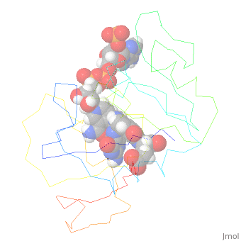
4p3r
From Proteopedia
(Difference between revisions)
| (One intermediate revision not shown.) | |||
| Line 1: | Line 1: | ||
| - | + | ||
==Cryogenic WT DHFR, time-averaged ensemble== | ==Cryogenic WT DHFR, time-averaged ensemble== | ||
| - | <StructureSection load='4p3r' size='340' side='right' caption='[[4p3r]], [[Resolution|resolution]] 1.15Å' scene=''> | + | <StructureSection load='4p3r' size='340' side='right'caption='[[4p3r]], [[Resolution|resolution]] 1.15Å' scene=''> |
== Structural highlights == | == Structural highlights == | ||
| - | <table><tr><td colspan='2'>[[4p3r]] is a 1 chain structure with sequence from [ | + | <table><tr><td colspan='2'>[[4p3r]] is a 1 chain structure with sequence from [https://en.wikipedia.org/wiki/Escherichia_coli_str._K-12_substr._DH10B Escherichia coli str. K-12 substr. DH10B]. Full crystallographic information is available from [http://oca.weizmann.ac.il/oca-bin/ocashort?id=4P3R OCA]. For a <b>guided tour on the structure components</b> use [https://proteopedia.org/fgij/fg.htm?mol=4P3R FirstGlance]. <br> |
| - | </td></tr><tr id=' | + | </td></tr><tr id='method'><td class="sblockLbl"><b>[[Empirical_models|Method:]]</b></td><td class="sblockDat" id="methodDat">X-ray diffraction, [[Resolution|Resolution]] 1.15Å</td></tr> |
| - | + | <tr id='ligand'><td class="sblockLbl"><b>[[Ligand|Ligands:]]</b></td><td class="sblockDat" id="ligandDat"><scene name='pdbligand=FOL:FOLIC+ACID'>FOL</scene>, <scene name='pdbligand=NAP:NADP+NICOTINAMIDE-ADENINE-DINUCLEOTIDE+PHOSPHATE'>NAP</scene></td></tr> | |
| - | <tr id=' | + | <tr id='resources'><td class="sblockLbl"><b>Resources:</b></td><td class="sblockDat"><span class='plainlinks'>[https://proteopedia.org/fgij/fg.htm?mol=4p3r FirstGlance], [http://oca.weizmann.ac.il/oca-bin/ocaids?id=4p3r OCA], [https://pdbe.org/4p3r PDBe], [https://www.rcsb.org/pdb/explore.do?structureId=4p3r RCSB], [https://www.ebi.ac.uk/pdbsum/4p3r PDBsum], [https://prosat.h-its.org/prosat/prosatexe?pdbcode=4p3r ProSAT]</span></td></tr> |
| - | + | ||
| - | <tr id='resources'><td class="sblockLbl"><b>Resources:</b></td><td class="sblockDat"><span class='plainlinks'>[ | + | |
</table> | </table> | ||
| - | {{Large structure}} | ||
| - | == Function == | ||
| - | [[http://www.uniprot.org/uniprot/B1XC49_ECODH B1XC49_ECODH]] Key enzyme in folate metabolism. Catalyzes an essential reaction for de novo glycine and purine synthesis, and for DNA precursor synthesis (By similarity).[PIRNR:PIRNR000194] | ||
<div style="background-color:#fffaf0;"> | <div style="background-color:#fffaf0;"> | ||
== Publication Abstract from PubMed == | == Publication Abstract from PubMed == | ||
| Line 22: | Line 17: | ||
</div> | </div> | ||
<div class="pdbe-citations 4p3r" style="background-color:#fffaf0;"></div> | <div class="pdbe-citations 4p3r" style="background-color:#fffaf0;"></div> | ||
| + | |||
| + | ==See Also== | ||
| + | *[[Dihydrofolate reductase 3D structures|Dihydrofolate reductase 3D structures]] | ||
== References == | == References == | ||
<references/> | <references/> | ||
__TOC__ | __TOC__ | ||
</StructureSection> | </StructureSection> | ||
| - | [[Category: | + | [[Category: Escherichia coli str. K-12 substr. DH10B]] |
| - | [[Category: | + | [[Category: Large Structures]] |
| - | + | [[Category: Fraser JS]] | |
| - | [[Category: Fraser | + | [[Category: Keedy DA]] |
| - | [[Category: Keedy | + | [[Category: Van den Bedem H]] |
| - | [[Category: | + | |
| - | + | ||
Current revision
Cryogenic WT DHFR, time-averaged ensemble
| |||||||||||