Sandbox Reserved 1387
From Proteopedia
(Difference between revisions)
| (18 intermediate revisions not shown.) | |||
| Line 2: | Line 2: | ||
==''Homo sapien'' Glucose Transporter GLUT3 / SLC2A3== | ==''Homo sapien'' Glucose Transporter GLUT3 / SLC2A3== | ||
<StructureSection load='5c65' size='400' side='right' caption='GLUT3' scene=''> | <StructureSection load='5c65' size='400' side='right' caption='GLUT3' scene=''> | ||
| - | + | __TOC__ | |
| - | + | ||
== Function == | == Function == | ||
The main function of this protein is glucose transmembrane transport. It is one of fourteen facilitative sugar transporters. <ref>https://www.ebi.ac.uk/pdbe/entry/pdb/5c65</ref> | The main function of this protein is glucose transmembrane transport. It is one of fourteen facilitative sugar transporters. <ref>https://www.ebi.ac.uk/pdbe/entry/pdb/5c65</ref> | ||
| - | GLUT3 is categorized as a Class I transporter due to its protein sequence and structural similarity to other glucose transporters grouped in Class I | + | GLUT3 is categorized as a Class I transporter due to its protein sequence and structural similarity to other glucose transporters grouped in Class I. GLUT3 displays the highest affinity for glucose of all of the Class I glucose transporters and has a transport capacity five times greater than that of GLUT1 and GLUT4<ref>DOI 10.1152/ajpendo.90388.2008</ref>. |
| + | |||
| + | GLUT3 is found predominantly in brain tissue, almost always expressed by neurons. The location of GLUT3 has led to it sometimes being called the "neuronal glucose transporter". | ||
| + | |||
| + | Its main function is to act in the transmembrane region to create channels for glucose to move across the cell membrane. First, the empty carrier opens to the cis side of the membrane for glucose to bind<ref>DOI 10.2147/CHC.S60484</ref>. Then the substrate binding carrier translocates to the trans side of the membrane where it then releases glucose on that side. Last the empty carrier switches to the cis side. | ||
== Disease == | == Disease == | ||
| - | Defects in GLUT3 can cause fetal death as well as neurodegeneration, which can lead to diseases like Alzheimer’s. | + | Defects in GLUT3 can cause fetal death as well as neurodegeneration, which can lead to diseases like Alzheimer’s. In the case of Alzheimer's Disease (AD), the defective GLUT3 does not provide neurons with optimal glucose concentrations. Autopsies of AD patients have shown higher sugar concentrations in the brain's extracellular tissue while also possessing abnormally low GLUT3 counts<ref>DOI 10.1016/j.jalz.2017.09.011</ref>. |
| + | |||
| + | Enhanced expression of GLUT3 is characteristic of several cancers, since this provides the rapidly dividing cells with a steady supply of glucose. Particularly, prostate cancer exhibits enhanced GLUT3 activity due to the presence of concentrated sugars in seminal fluid<ref>DOI 10.1002/ijc.31165</ref>. | ||
| - | + | Finally, Type 2 Diabetes patients exhibit a decrease in the amount of transcripts that encode for GLUT3. Because they lack the appropriate amount of GLUT3, sugars will accumulate on the outside of diabetic cells. | |
== Structural highlights == | == Structural highlights == | ||
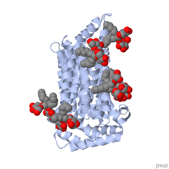
| - | < | + | GLUT3 has 481 amino acids, which compose 28 alpha helices. It weighs approximately 52,520 Daltons which is equivalent to 8.72115e-20 grams<ref>https://www.ebi.ac.uk/pdbe/entry/pdb/5c65</ref>. |
| - | <scene name='77/777707/Hydrophilic_binding_sites/1'>Hydrophilic Binding Sites</scene> | + | Its <scene name='77/777707/Hydrophobic_binding_sites/2'>Hydrophobic Binding Sites</scene> are Leucine, Alanine, Phenylalanine, Isoleucine. Its <scene name='77/777707/Hydrophilic_binding_sites/1'>Hydrophilic Binding Sites</scene> are Serine, Glutamic Acid, Arginine, Threonine. The interactions between the hydrophobic and hydrophylic groups allow GLUT3 to bind to octylglucose neopentylglycol and cholesterol hemisuccinate. Both of these substrates have polar and non-polar regions; they are precursors to simpler sugars. |
| - | + | The protein's <scene name='77/777707/Non_polar_scene/1'>Non Polar Regions</scene> are understandably located on the exterior surface, since the complex is embedded in the lipid membrane. Conversely, GLUT3's <scene name='77/777707/Polar_regions/1'>Polar Regions</scene> are located on the inside of the complex to interact with the substrates and hold the structure of the protein together. | |
</StructureSection> | </StructureSection> | ||
== References == | == References == | ||
<references/> | <references/> | ||
Current revision
| This Sandbox is Reserved from January through July 31, 2018 for use in the course HLSC322: Principles of Genetics and Genomics taught by Genevieve Houston-Ludlam at the University of Maryland, College Park, USA. This reservation includes Sandbox Reserved 1311 through Sandbox Reserved 1430. |
To get started:
More help: Help:Editing |
Homo sapien Glucose Transporter GLUT3 / SLC2A3
| |||||||||||
References
- ↑ https://www.ebi.ac.uk/pdbe/entry/pdb/5c65
- ↑ Simpson IA, Dwyer D, Malide D, Moley KH, Travis A, Vannucci SJ. The facilitative glucose transporter GLUT3: 20 years of distinction. Am J Physiol Endocrinol Metab. 2008 Aug;295(2):E242-53. doi:, 10.1152/ajpendo.90388.2008. Epub 2008 Jun 24. PMID:18577699 doi:http://dx.doi.org/10.1152/ajpendo.90388.2008
- ↑ doi: https://dx.doi.org/10.2147/CHC.S60484
- ↑ An Y, Varma VR, Varma S, Casanova R, Dammer E, Pletnikova O, Chia CW, Egan JM, Ferrucci L, Troncoso J, Levey AI, Lah J, Seyfried NT, Legido-Quigley C, O'Brien R, Thambisetty M. Evidence for brain glucose dysregulation in Alzheimer's disease. Alzheimers Dement. 2017 Oct 19. pii: S1552-5260(17)33765-2. doi:, 10.1016/j.jalz.2017.09.011. PMID:29055815 doi:http://dx.doi.org/10.1016/j.jalz.2017.09.011
- ↑ Gonzalez-Menendez P, Hevia D, Mayo JC, Sainz RM. The dark side of glucose transporters in prostate cancer: Are they a new feature to characterize carcinomas? Int J Cancer. 2017 Nov 21. doi: 10.1002/ijc.31165. PMID:29159872 doi:http://dx.doi.org/10.1002/ijc.31165
- ↑ https://www.ebi.ac.uk/pdbe/entry/pdb/5c65
