This old version of Proteopedia is provided for student assignments while the new version is undergoing repairs. Content and edits done in this old version of Proteopedia after March 1, 2026 will eventually be lost when it is retired in about June of 2026.
Apply for new accounts at the new Proteopedia. Your logins will work in both the old and new versions.
User:Karsten Theis/Takustr6
From Proteopedia
(→Introduction) |
|||
| (16 intermediate revisions not shown.) | |||
| Line 7: | Line 7: | ||
==Results== | ==Results== | ||
| - | <StructureSection load='' size=' | + | <StructureSection load='' size='500' side='right' caption='Saenger lab structures' scene='79/793353/2bj6/1'> |
=== 1973 === | === 1973 === | ||
| - | <scene name='79/793353/Aharfu01/ | + | <scene name='79/793353/Aharfu01/2'> |
O2,2'-Cyclouridine</scene> (CSD AHARFU01) | O2,2'-Cyclouridine</scene> (CSD AHARFU01) | ||
| Line 29: | Line 29: | ||
=== 1988 - 1993 === | === 1988 - 1993 === | ||
| - | Proteinase K (2PRK) | + | Proteinase K (<scene name='79/793353/2prk/1'>2PRK</scene>) |
... as well as 1PEK and 1PTK | ... as well as 1PEK and 1PTK | ||
| Line 35: | Line 35: | ||
=== 1992 === | === 1992 === | ||
| - | Factor for inversion stimulation FIS (1FIA) | + | Factor for inversion stimulation FIS (<scene name='79/793353/2prk/2'>1FIA</scene>) |
=== 1994 - 2012 === | === 1994 - 2012 === | ||
| - | Tetracycline repressor ( | + | <jmol> |
| + | <jmolLink> | ||
| + | <script> load files "=2TRT" "=2TRT" "=1QPI" "=1QPI" filter "biomolecule 1" | ||
| + | compare {3.1} {1.1} SUBSET{*.CA} ATOMS{8-155}{8-155} ROTATE TRANSLATE | ||
| + | compare {4.1} {1.1} SUBSET{*.CA} ATOMS{8-155}{8-155} ROTATE TRANSLATE | ||
| + | set refreshing FALSE | ||
| + | moveto /* time, axisAngle */ 1.0 { 564 643 518 109.44} /* zoom, translation */ 110.0 0.0 0.0 /* center, rotationRadius */ {34.160000000000075 34.16000000000002 25.835952990815013} 46.11980702931616 /* navigation center, translation, depth */ {0 0 0} 0 0 0 /* cameraDepth, cameraX, cameraY */ 3.0 0.0 0.0; | ||
| + | restrict none | ||
| + | select 1.1 and (TAC or _MG) | ||
| + | spacefill on | ||
| + | select protein; backbone 0.3; color gray | ||
| + | select 4.1 and DNA; cartoon only; color orange | ||
| + | select 27-44 and protein; cartoon only; color skyblue | ||
| + | animation fps 1 | ||
| + | animation mode loop | ||
| + | animation FRAMES [1 2 3 4 3 2] | ||
| + | set refreshing TRUE | ||
| + | animation on | ||
| + | </script> | ||
| + | <text>Tetracycline repressor</text> | ||
| + | </jmolLink> | ||
| + | </jmol> switching back an forth between the tetracycline bound form (does not bind to DNA) and the DNA-bound form (no tetracycline bound. The two structures with neither DNA nor tetracycline bound are hypothetical and are shown to better illustrate the conformational change between the two complexes. | ||
| + | |||
| + | <jmol> | ||
| + | <jmolRadioGroup> | ||
| + | <item> | ||
| + | <script>animation off; delay 1.0; model 1</script> | ||
| + | <text>Tetracycline complex</text> | ||
| + | </item> | ||
| + | <item> | ||
| + | <script>animation off; delay 1.0; model 4</script> | ||
| + | <text>DNA complex</text> | ||
| + | </item> | ||
| + | <item> | ||
| + | <script>animation off; delay 1.0; model 2</script> | ||
| + | <text>Tetracycline hidden</text> | ||
| + | </item> | ||
| + | <item> | ||
| + | <script>animation off; delay 1.0; model 3</script> | ||
| + | <text>DNA hidden</text> | ||
| + | </item> | ||
| + | <item> | ||
| + | <script>animation on</script> | ||
| + | <text>animations</text> | ||
| + | <checked>true</checked> | ||
| + | |||
| + | </item> | ||
| + | |||
| + | </jmolRadioGroup> | ||
| + | </jmol> | ||
| + | |||
=== 1997 === | === 1997 === | ||
| Line 46: | Line 96: | ||
=== 1998 === | === 1998 === | ||
| + | <scene name='79/793353/1auk/2'> | ||
| + | Arylsulfatase A</scene> (1AUK) | ||
| + | |||
| + | ::"This was a side project for me just after I graduated from the lab working on the biochemistry of the DNA bending protein FIS. I was hanging around in the lab looking for a postdoctoral position, and a new graduate student, Gordian Lukatela, visited bringing crystals of the human lysosomal protein arylsulfatase A. We made good progress, but the project got delayed twice. First, the antibody-affinity column used in purification failed, and we lost our supply of fresh protein. Second, a nasty bug in data processing compromised our structure factors, leading to abnormally high R-factors for a structure that was essentially correct. | ||
| - | + | ::The resulting structure is fascinating because of its unusual modification in the active site with an amino acid side chain carrying an aldehyde functional group. In the crystal structure, the density looked like the aldehyde was hydrated (geminal diol), but we weren't bold enough to commit to that (at least not for the pdb - there is a figure with the density and interpretation in the paper). However, we came up with a mechanism that utilized this hydrated aldehyde. A higher resolution structure of a bacterial homolog (1hdh, solved three years later in 2001) showed the hydrated aldehyde as the resting state of the active site, supporting our mechanistic hypothesis." (Karsten) | |
=== 1996 - 2001<ref>Karsten: Looking back, I'm amazed that only five years passed between the 4 Angstrom model and the complete atomic model of photosystem I. To me, folks working on that project were in a different space-time continuum. Everything took longer, the unit cell dimensions were crazy, the solvent content unbelievable, and then the did these things like advancing the spindle every three images because they were burning through the crystal, or using a cylindrical image plate in Japan in hopes of spreading out the overlapping diffraction spots. And then there was airport security: "No, you can't X-ray that, sorry. It's in the thermos to keep it cool. No, you can't look inside, it is light sensitive. Yes, trust me." I am still impressed by the graduate students who dared to join this project.</ref> === | === 1996 - 2001<ref>Karsten: Looking back, I'm amazed that only five years passed between the 4 Angstrom model and the complete atomic model of photosystem I. To me, folks working on that project were in a different space-time continuum. Everything took longer, the unit cell dimensions were crazy, the solvent content unbelievable, and then the did these things like advancing the spindle every three images because they were burning through the crystal, or using a cylindrical image plate in Japan in hopes of spreading out the overlapping diffraction spots. And then there was airport security: "No, you can't X-ray that, sorry. It's in the thermos to keep it cool. No, you can't look inside, it is light sensitive. Yes, trust me." I am still impressed by the graduate students who dared to join this project.</ref> === | ||
| - | Photosystem I ( | + | Photosystem I (1jb0) |
| + | This is a big protein and takes a while to load. The <scene name='79/793353/Psi_overall/1'>figure</scene> shows a single monomer of the trimeric structure. | ||
| + | |||
| + | Exciton transfer: If you press the buttons, you see animations of a chlorophyll molecule getting excited by a flash of light, and transferring the energy through exciton transfer. PSI has a built in antenna system, transferring excitation energy by exciton transfer to the special pair, where the redox chemistry (charge separation) is initiated. Exciton transfer is a stochastic process so every exciton transfer pathway is different. There are two buttons to show two possiblities It's not entirely correct, but it looks pretty (inspired by the famous cytochrome picture by Irving Geis[https://www.l2molecule.com/inspirations/2015/1/24/irving-geis-and-his-paintings-of-proteins]) | ||
| + | |||
| + | <jmol> | ||
| + | <jmolButton> | ||
| + | <script> script "http://proteopedia.org/wiki/images/c/c2/Psi_animation2.spt" | ||
| + | </script> | ||
| + | <text>random path 1</text> | ||
| + | </jmolButton> | ||
| + | </jmol><jmol> | ||
| + | <jmolButton> | ||
| + | <script> script "http://proteopedia.org/wiki/images/9/90/Psi_animation1.spt" | ||
| + | </script> | ||
| + | <text>random path 2</text> | ||
| + | </jmolButton> | ||
| + | </jmol> | ||
| + | |||
| + | |||
| + | Electron transfer: Once one of the special pair chlorophylls is excited, charge separation and electron transfer take place. This <scene name='79/793353/Psi_overall/3'>figure</scene> shows the cofactors involved in transferring electrons. | ||
=== 1999 === | === 1999 === | ||
| - | Cycloamylose 26 1C58 | + | Cycloamylose 26 <scene name='79/793353/1c58/2'>1C58</scene> |
| Line 90: | Line 164: | ||
| + | === 2017 === | ||
| + | |||
| + | <scene name='79/793353/5jiw/1'>Amylomaltase</scene> (5JIW) | ||
| + | |||
| + | There is a spectacular length of <scene name='79/793353/5jiw/2'>amylose</scene> in this structure... | ||
</StructureSection> | </StructureSection> | ||
| Line 110: | Line 189: | ||
=== Structure refinement === | === Structure refinement === | ||
1992: For every new structure, there would be two papers. The first describing the fold (in Science or Nature), the second "full paper" describing the details after refining the structure. Then, if you wanted, you would deposit your coordinates in the PDB. The diffraction data would be moved to magnetic tape as soon as a new project took up to much of your precious disk space quota. | 1992: For every new structure, there would be two papers. The first describing the fold (in Science or Nature), the second "full paper" describing the details after refining the structure. Then, if you wanted, you would deposit your coordinates in the PDB. The diffraction data would be moved to magnetic tape as soon as a new project took up to much of your precious disk space quota. | ||
| + | |||
| + | ===Theoretical and computational work === | ||
=== Making figures === | === Making figures === | ||
| Line 124: | Line 205: | ||
== Discussion == | == Discussion == | ||
| - | + | see discussion page, second tab on the top | |
| - | + | ||
| - | + | ||
| - | + | ||
| - | + | ||
| - | + | ||
| - | + | ||
| - | + | ||
| - | + | ||
| - | + | ||
| - | + | ||
| - | + | ||
| - | + | ||
| - | + | ||
| - | + | ||
| - | + | ||
| - | + | ||
| - | + | ||
| - | + | ||
| - | + | ||
| - | + | ||
| - | + | ||
| - | + | ||
| - | + | ||
| - | + | ||
| - | + | ||
| - | + | ||
| - | + | ||
| - | + | ||
| - | + | ||
| - | + | ||
| - | + | ||
| - | + | ||
| - | + | ||
| - | + | ||
| - | + | ||
| - | + | ||
| - | + | ||
| - | + | ||
| - | + | ||
| - | + | ||
| - | + | ||
| - | + | ||
== Footnotes == | == Footnotes == | ||
<references/> | <references/> | ||
Current revision
Introduction
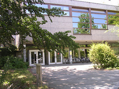
The Institute of Crystallography, part of the Chemistry department of the Free University Berlin, was located at Takustrasse 6. The macromolecular crystallography was on the third floor. It is still there, but the institutional framework has changed, and a new group has moved in. This page reminisces about research done from when the group moved to Berlin to the PIs retirement.
Results
| |||||||||||
Methods
The third floor was one big corridor, outlining the steps in structure solution. Cold rooms on one end, crystallization and X-ray labs in the middle (along with the two grown-up offices and the seminar room) and data crunching on the other end.
Purification
Crystallization
In-house data collection
Synchrotron data collection
Data processing
Structure solution
Structure refinement
1992: For every new structure, there would be two papers. The first describing the fold (in Science or Nature), the second "full paper" describing the details after refining the structure. Then, if you wanted, you would deposit your coordinates in the PDB. The diffraction data would be moved to magnetic tape as soon as a new project took up to much of your precious disk space quota.
Theoretical and computational work
Making figures
1992: FTP (yes, that's a verb) your coordinates to the Evans-and-Sutherland vector graphic machine (sign up first). Find the perfect orientation of your model (in stereo, if necessary), apply rub-off lettering directly on the screen for labels, and set up a tripod for the camera to take pictures. If you were in a hurry, run down to the photography lab in the basement to develop your film and make copies of the negatives (unless you were making slides for a talk). Then, patiently take off all the lettering and start over because someone wanted a different view, maybe 3 degrees rotation around the y-axis.
Publishing
1992: We had computers, even PCs, but they were slow. You would find out if you had your entire PhD thesis in a single document and tried to go from chapter 1 to chapter 4 after making changes. Word had to re-paginate the entire document, which would take longer than making a pot of coffee and drinking it. Others used LaTeX on the mainframe, entering text on a VT100 terminal.
Model building
1990s: Every time a new structure was solved, we would build a physical model. Well, we would have a crystallography course, and participants would build stretches of ten amino acids from a list of main-chain and side-chain torsion angles. Then, we would assemble those on a wooden board with aluminum stakes, guided by the projection of the C-alpha trace taped to the wooden board. At the end, the technician from downstairs would build a plexiglass housing to avoid tampering with the structure (see Discussion, occupational hazards). It was reminiscent of a hunter putting skulls and antlers of the kill on the wall.
Conferences
Discussion
see discussion page, second tab on the top
Footnotes
- ↑ Karsten: back then, you could ask for a specific PDB ID, and researchers tried to get one that fit their project. nRNT for RNase T1 is pretty good, but at one point the lab ran out of digits
- ↑ Karsten: Looking back, I'm amazed that only five years passed between the 4 Angstrom model and the complete atomic model of photosystem I. To me, folks working on that project were in a different space-time continuum. Everything took longer, the unit cell dimensions were crazy, the solvent content unbelievable, and then the did these things like advancing the spindle every three images because they were burning through the crystal, or using a cylindrical image plate in Japan in hopes of spreading out the overlapping diffraction spots. And then there was airport security: "No, you can't X-ray that, sorry. It's in the thermos to keep it cool. No, you can't look inside, it is light sensitive. Yes, trust me." I am still impressed by the graduate students who dared to join this project.
