Journal:Acta Cryst D:S2059798319000214
From Proteopedia
(Difference between revisions)

(New page: -) |
|||
| (32 intermediate revisions not shown.) | |||
| Line 1: | Line 1: | ||
| - | - | + | <StructureSection load='' size='450' side='right' scene='80/805753/Cv/2' caption=''> |
| + | ===In-house high energy remote SAD-phasing using the magic triangle: how to tackle the P1 low symmetry using multiple orientations on the same human IBA57 crystal to increase multiplicity=== | ||
| + | <big>Spyridon Gourdoupis, Veronica Nasta, Simone Ciofi-Baffoni, Lucia Banci and Vito Calderone </big> <ref>doi 10.1107/S2059798319000214</ref> | ||
| + | <hr/> | ||
| + | <b>Molecular Tour</b><br> | ||
| + | IBA57 is a 325-residue human protein which localizes to the mitochondrion and is part of the iron-sulfur cluster assembly pathway <ref name="Rouault">PMID:25425402</ref>, <ref name="Andreini">PMID:28135316</ref>. | ||
| + | The maturation of mitochondrial iron-sulfur proteins requires a complex protein machinery. In the late steps of this machinery, a [2Fe-2S] cluster is converted into a [4Fe-4S] cluster. Human IBA57 protein acts in this step as an iron-sulfur cluster assembly component along with ISCA1 and ISCA2 <ref name="Mikolajczyk">PMID:25347204</ref>, <ref name="Brancaccio">PMID:27989128</ref>, <ref name="Ciofi-Baffoni">PMID:29219157</ref>, <ref name="Gourdoupis">PMID:30269484</ref>. | ||
| + | The structure of this protein was still unknown and the closest homologue whose structure was determined showed 25% sequence homology only (which made molecular replacement in principle unlikely to be successful). For this reason, experimental phasing was the most likely way to solve it. | ||
| + | |||
| + | This paper (DOI 10.1107/S2059798319000214) describes the approach used to solve in-house the structure of human IBA57 through 5-amino-2,4,6-triiodoisophthalic acid (I3C) high energy remote SAD-phasing. Multiple orientations (each of them corresponding to a different run) of the same P1 (triclinic) crystal have been exploited to acquire sufficient real data multiplicity for successful phasing and thus minimizing the difficulties of merging datasets coming from different crystals. | ||
| + | |||
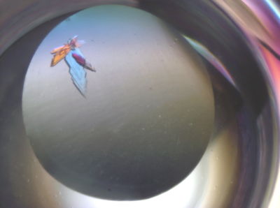
| + | [[Image:IBA57a.jpg|left|400px|thumb|Screenshot of IBA57 crystals]] | ||
| + | {{Clear}} | ||
| + | |||
| + | It is also described how the joint use of this I3C derivative and of an in-house native dataset through a SIRAS approach decreases the data multiplicity needed for phasing by almost 50%. | ||
| + | |||
| + | Furthermore, it is illustrated that there is a clear data multiplicity threshold value for success and failure in phasing and how adding further data does not significantly affect substructure solution and model building. The multiplicity threshold for successful phasing appeared, in fact, to be around five, independent of the combination of datasets (runs) used. This value can be reduced to about half through SIRAS by exploiting the isomorphous differences with a second native dataset reaching a successful multiplicity value of less than three. | ||
| + | |||
| + | To our knowledge, this is the only structure present in the PDB which has been solved in-house, through remote SAD, in space group P1 and using one crystal only. All the raw data used, deriving from the different orientations (runs), have been deposited to Zenodo (DOI 10.5281/zenodo.2531553)<ref>doi 10.5281/zenodo.2531553</ref> both for educational purposes and to enable other crystallographers to improve methods for data processing and structure solution and thus to benefit from these findings. | ||
| + | At the time of structure solution coordinates and structure factors were deposited and released in the Protein Data Bank under the accession codes [[5oli]] (for the in-house I3C derivative) and [[6esr]] (for the higher resolution synchrotron structure); at the time of writing the manuscript both entries have been re-refined in order to optimize model quality and statistics and so they have been superseded by [[6qe4]] and [[6qe3]] respectively. | ||
| + | |||
| + | <scene name='80/805753/Cv/6'>Secondary structure ribbon representation of the structure of human IBA57</scene> with the residues for which even the main chain electron density is very poor if not absent at all highlighted in red (53-59, 61, 88-92, 115-118, 138-147, 262, 296-300, 306-311). | ||
| + | |||
| + | <scene name='80/805753/Cv/7'>Superposition</scene> between [[6esr]] (red) and [[5oli]] (green) secondary structures. It appears that, in the case of [[6esr]], there is a slight loss in secondary structure elements (mainly β-strands in the N-terminus region) with respect to [[5oli]] and there is the appearance of a very short 3/10 helix around residue 90. It must be pointed out anyway that those regions mostly correspond to the regions in which electron density is very weak and thus model tracing can be quite approximate. | ||
| + | |||
| + | *<scene name='80/805753/Cv/9'>1st I3C binding site</scene>. Water molecules are shown as red spheres. | ||
| + | *<scene name='80/805753/Cv/11'>2nd I3C binding site</scene>. | ||
| + | *<scene name='80/805753/Cv/12'>3th I3C binding site</scene>. | ||
| + | *<scene name='80/805753/Cv/14'>4th I3C binding site</scene>. | ||
| + | |||
| + | '''PDB references:''' Re-refinement of 6ESR human IBA57 at 1.75 A resolution [[6qe3]]; Re-refinement of 5OLI human IBA57-I3C [[6qe4]]. | ||
| + | |||
| + | <b>References</b><br> | ||
| + | <references/> | ||
| + | </StructureSection> | ||
| + | __NOEDITSECTION__ | ||
Current revision
| |||||||||||
This page complements a publication in scientific journals and is one of the Proteopedia's Interactive 3D Complement pages. For aditional details please see I3DC.

