Journal:Acta Cryst D:S2059798319000214
From Proteopedia
(Difference between revisions)

| (6 intermediate revisions not shown.) | |||
| Line 10: | Line 10: | ||
This paper (DOI 10.1107/S2059798319000214) describes the approach used to solve in-house the structure of human IBA57 through 5-amino-2,4,6-triiodoisophthalic acid (I3C) high energy remote SAD-phasing. Multiple orientations (each of them corresponding to a different run) of the same P1 (triclinic) crystal have been exploited to acquire sufficient real data multiplicity for successful phasing and thus minimizing the difficulties of merging datasets coming from different crystals. | This paper (DOI 10.1107/S2059798319000214) describes the approach used to solve in-house the structure of human IBA57 through 5-amino-2,4,6-triiodoisophthalic acid (I3C) high energy remote SAD-phasing. Multiple orientations (each of them corresponding to a different run) of the same P1 (triclinic) crystal have been exploited to acquire sufficient real data multiplicity for successful phasing and thus minimizing the difficulties of merging datasets coming from different crystals. | ||
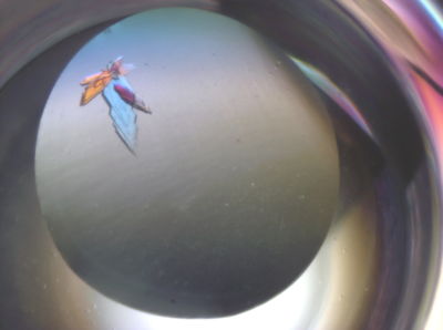
| - | [[Image:IBA57a.jpg|left| | + | [[Image:IBA57a.jpg|left|400px|thumb|Screenshot of IBA57 crystals]] |
{{Clear}} | {{Clear}} | ||
| Line 22: | Line 22: | ||
<scene name='80/805753/Cv/6'>Secondary structure ribbon representation of the structure of human IBA57</scene> with the residues for which even the main chain electron density is very poor if not absent at all highlighted in red (53-59, 61, 88-92, 115-118, 138-147, 262, 296-300, 306-311). | <scene name='80/805753/Cv/6'>Secondary structure ribbon representation of the structure of human IBA57</scene> with the residues for which even the main chain electron density is very poor if not absent at all highlighted in red (53-59, 61, 88-92, 115-118, 138-147, 262, 296-300, 306-311). | ||
| - | <scene name='80/805753/Cv/7'>Superposition</scene> between [[6esr]] (red) and [[5oli]] (green) secondary structures. It appears that, | + | <scene name='80/805753/Cv/7'>Superposition</scene> between [[6esr]] (red) and [[5oli]] (green) secondary structures. It appears that, in the case of [[6esr]], there is a slight loss in secondary structure elements (mainly β-strands in the N-terminus region) with respect to [[5oli]] and there is the appearance of a very short 3/10 helix around residue 90. It must be pointed out anyway that those regions mostly correspond to the regions in which electron density is very weak and thus model tracing can be quite approximate. |
| - | in the case of [[6esr]], there is a slight loss in secondary structure elements (mainly β-strands in the N-terminus region) with respect to [[5oli]] and there is the appearance of a very short 3/10 helix around residue 90. It must be pointed out anyway that those regions mostly correspond to the regions in which electron density is very weak and thus model tracing can be quite approximate. | + | |
| + | *<scene name='80/805753/Cv/9'>1st I3C binding site</scene>. Water molecules are shown as red spheres. | ||
| + | *<scene name='80/805753/Cv/11'>2nd I3C binding site</scene>. | ||
| + | *<scene name='80/805753/Cv/12'>3th I3C binding site</scene>. | ||
| + | *<scene name='80/805753/Cv/14'>4th I3C binding site</scene>. | ||
| + | |||
| + | '''PDB references:''' Re-refinement of 6ESR human IBA57 at 1.75 A resolution [[6qe3]]; Re-refinement of 5OLI human IBA57-I3C [[6qe4]]. | ||
<b>References</b><br> | <b>References</b><br> | ||
Current revision
| |||||||||||
This page complements a publication in scientific journals and is one of the Proteopedia's Interactive 3D Complement pages. For aditional details please see I3DC.

