This old version of Proteopedia is provided for student assignments while the new version is undergoing repairs. Content and edits done in this old version of Proteopedia after March 1, 2026 will eventually be lost when it is retired in about June of 2026.
Apply for new accounts at the new Proteopedia. Your logins will work in both the old and new versions.
1cii
From Proteopedia
(Difference between revisions)
| (One intermediate revision not shown.) | |||
| Line 3: | Line 3: | ||
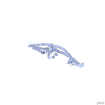
<StructureSection load='1cii' size='340' side='right'caption='[[1cii]], [[Resolution|resolution]] 3.00Å' scene=''> | <StructureSection load='1cii' size='340' side='right'caption='[[1cii]], [[Resolution|resolution]] 3.00Å' scene=''> | ||
== Structural highlights == | == Structural highlights == | ||
| - | <table><tr><td colspan='2'>[[1cii]] is a 1 chain structure. Full crystallographic information is available from [http://oca.weizmann.ac.il/oca-bin/ocashort?id=1CII OCA]. For a <b>guided tour on the structure components</b> use [ | + | <table><tr><td colspan='2'>[[1cii]] is a 1 chain structure with sequence from [https://en.wikipedia.org/wiki/Escherichia_coli Escherichia coli]. Full crystallographic information is available from [http://oca.weizmann.ac.il/oca-bin/ocashort?id=1CII OCA]. For a <b>guided tour on the structure components</b> use [https://proteopedia.org/fgij/fg.htm?mol=1CII FirstGlance]. <br> |
| - | </td></tr><tr id='resources'><td class="sblockLbl"><b>Resources:</b></td><td class="sblockDat"><span class='plainlinks'>[ | + | </td></tr><tr id='method'><td class="sblockLbl"><b>[[Empirical_models|Method:]]</b></td><td class="sblockDat" id="methodDat">X-ray diffraction, [[Resolution|Resolution]] 3Å</td></tr> |
| + | <tr id='resources'><td class="sblockLbl"><b>Resources:</b></td><td class="sblockDat"><span class='plainlinks'>[https://proteopedia.org/fgij/fg.htm?mol=1cii FirstGlance], [http://oca.weizmann.ac.il/oca-bin/ocaids?id=1cii OCA], [https://pdbe.org/1cii PDBe], [https://www.rcsb.org/pdb/explore.do?structureId=1cii RCSB], [https://www.ebi.ac.uk/pdbsum/1cii PDBsum], [https://prosat.h-its.org/prosat/prosatexe?pdbcode=1cii ProSAT]</span></td></tr> | ||
</table> | </table> | ||
== Function == | == Function == | ||
| - | [ | + | [https://www.uniprot.org/uniprot/CEIA_ECOLX CEIA_ECOLX] This colicin is a channel-forming colicin. This class of transmembrane toxins depolarize the cytoplasmic membrane, leading to dissipation of cellular energy. Colicins are polypeptide toxins produced by and active against E.coli and closely related bacteria. |
== Evolutionary Conservation == | == Evolutionary Conservation == | ||
[[Image:Consurf_key_small.gif|200px|right]] | [[Image:Consurf_key_small.gif|200px|right]] | ||
| Line 18: | Line 19: | ||
</jmol>, as determined by [http://consurfdb.tau.ac.il/ ConSurfDB]. You may read the [[Conservation%2C_Evolutionary|explanation]] of the method and the full data available from [http://bental.tau.ac.il/new_ConSurfDB/main_output.php?pdb_ID=1cii ConSurf]. | </jmol>, as determined by [http://consurfdb.tau.ac.il/ ConSurfDB]. You may read the [[Conservation%2C_Evolutionary|explanation]] of the method and the full data available from [http://bental.tau.ac.il/new_ConSurfDB/main_output.php?pdb_ID=1cii ConSurf]. | ||
<div style="clear:both"></div> | <div style="clear:both"></div> | ||
| - | <div style="background-color:#fffaf0;"> | ||
| - | == Publication Abstract from PubMed == | ||
| - | The ion-channel forming colicins A, B, E1, Ia, Ib and N all kill bacterial cells selectively by co-opting bacterial active-transport pathways and forming voltage-gated ion conducting channels across the plasma membrane of the target bacterium. The crystal structure of colicin Ia reveals a molecule 210 A long with three distinct functional domains arranged along a backbone of two extraordinarily long alpha-helices. A central domain at the bend of the hairpin-like structure mediates specific recognition and binding to an outer-membrane receptor. A second domain mediates translocation across the outer membrane via the TonB transport pathway; the TonB-box recognition element of colicin Ia is on one side of three 80 A-long helices arranged as a helical sheet. A third domain is made up of 10 alpha-helices which form a voltage-activated and voltage-gated ion conducting channel across the plasma membrane of the target cell. The two 160 A-long alpha-helices that link the receptor-binding domain to the other domains enable the colicin Ia molecule to span the periplasmic space and contact both the outer and plasma membranes simultaneously during function. | ||
| - | |||
| - | Crystal structure of colicin Ia.,Wiener M, Freymann D, Ghosh P, Stroud RM Nature. 1997 Jan 30;385(6615):461-4. PMID:9009197<ref>PMID:9009197</ref> | ||
| - | |||
| - | From MEDLINE®/PubMed®, a database of the U.S. National Library of Medicine.<br> | ||
| - | </div> | ||
| - | <div class="pdbe-citations 1cii" style="background-color:#fffaf0;"></div> | ||
==See Also== | ==See Also== | ||
*[[Colicin|Colicin]] | *[[Colicin|Colicin]] | ||
*[[Colicin 3D structures|Colicin 3D structures]] | *[[Colicin 3D structures|Colicin 3D structures]] | ||
| - | *[[Colicin Ia|Colicin Ia]] | ||
| - | == References == | ||
| - | <references/> | ||
__TOC__ | __TOC__ | ||
</StructureSection> | </StructureSection> | ||
| + | [[Category: Escherichia coli]] | ||
[[Category: Large Structures]] | [[Category: Large Structures]] | ||
| - | [[Category: Freymann | + | [[Category: Freymann D]] |
| - | [[Category: Ghosh | + | [[Category: Ghosh P]] |
| - | [[Category: Stroud | + | [[Category: Stroud R]] |
| - | [[Category: Wiener | + | [[Category: Wiener M]] |
| - | + | ||
| - | + | ||
| - | + | ||
| - | + | ||
Current revision
COLICIN IA
| |||||||||||