We apologize for Proteopedia being slow to respond. For the past two years, a new implementation of Proteopedia has been being built. Soon, it will replace this 18-year old system. All existing content will be moved to the new system at a date that will be announced here.
Sandbox Reserved 1620
From Proteopedia
(Difference between revisions)
| (2 intermediate revisions not shown.) | |||
| Line 8: | Line 8: | ||
The [https://en.wikipedia.org/wiki/ABCG2 ABCG2 multidrug transporter] is a membrane protein from the '''A'''TP-'''B'''inding '''C'''assette [https://en.wikipedia.org/wiki/ATP-binding_cassette_transporter (ABC)] transporter family, specifically the G-subfamily. Also known as the breast cancer resistance protein (BCRP), ABCG2 has physiological roles in various tissue cells including the [https://en.wikipedia.org/wiki/Mammary_gland mammary gland] and the [https://en.wikipedia.org/wiki/Blood%E2%80%93brain_barrier blood-brain], [https://en.wikipedia.org/wiki/Blood%E2%80%93testis_barrier blood-testis], and [https://en.wikipedia.org/wiki/Placenta maternal-fetal] barriers.<ref name="Taylor">PMID:28554189</ref> ABCG2 protects cells by exporting [https://en.wikipedia.org/wiki/Xenobiotic xenobiotic] molecules out of the cell using ATP hydrolysis. ABCG2 also affects the [https://en.wikipedia.org/wiki/Pharmacokinetics pharmacokinetics] of many drugs and contributes to [https://en.wikipedia.org/wiki/Multiple_drug_resistance multidrug resistance].<ref name="Manolaridis">PMID:30405239</ref> | The [https://en.wikipedia.org/wiki/ABCG2 ABCG2 multidrug transporter] is a membrane protein from the '''A'''TP-'''B'''inding '''C'''assette [https://en.wikipedia.org/wiki/ATP-binding_cassette_transporter (ABC)] transporter family, specifically the G-subfamily. Also known as the breast cancer resistance protein (BCRP), ABCG2 has physiological roles in various tissue cells including the [https://en.wikipedia.org/wiki/Mammary_gland mammary gland] and the [https://en.wikipedia.org/wiki/Blood%E2%80%93brain_barrier blood-brain], [https://en.wikipedia.org/wiki/Blood%E2%80%93testis_barrier blood-testis], and [https://en.wikipedia.org/wiki/Placenta maternal-fetal] barriers.<ref name="Taylor">PMID:28554189</ref> ABCG2 protects cells by exporting [https://en.wikipedia.org/wiki/Xenobiotic xenobiotic] molecules out of the cell using ATP hydrolysis. ABCG2 also affects the [https://en.wikipedia.org/wiki/Pharmacokinetics pharmacokinetics] of many drugs and contributes to [https://en.wikipedia.org/wiki/Multiple_drug_resistance multidrug resistance].<ref name="Manolaridis">PMID:30405239</ref> | ||
| - | ABCG2 belongs to the family of 48 transporter proteins called ATP-binding cassette transporters (ABC transporters). The ABC transporters differ from each other by their size structure, and ordering of domains. A high percentage of the ABC family transporters (19 of the 48) transport [https://en.wikipedia.org/wiki/List_of_chemotherapeutic_agents chemotherapeutic agents] out of cells, making expression levels of ABC transporters a major indicator of cancer treatment prognosis.<ref name="Robey">PMID:29643473</ref> Using [https://en.wikipedia.org/wiki/Cryogenic_electron_microscopy cryoelectron microscopy] (Cryo EM), the two cavity substrate transport structure (<scene name='83/832932/Highlight_cavity_1/3'>Cavity 1</scene>; <scene name='83/832932/Atp_bound_use_cav_2/3'>Cavity 2</scene>), inward facing nucleotide binding domain (<scene name='83/832932/Overall_structure_nbd_unbound/6'>NBD</scene>), and condensed extracellular loop 3 (<scene name='83/832939/El-3/ | + | ABCG2 belongs to the family of 48 transporter proteins called ATP-binding cassette transporters (ABC transporters). The ABC transporters differ from each other by their size, structure, and ordering of domains. A high percentage of the ABC family transporters (19 of the 48) transport [https://en.wikipedia.org/wiki/List_of_chemotherapeutic_agents chemotherapeutic agents] out of cells, making expression levels of ABC transporters a major indicator of cancer treatment prognosis.<ref name="Robey">PMID:29643473</ref> Using [https://en.wikipedia.org/wiki/Cryogenic_electron_microscopy cryoelectron microscopy] (Cryo EM), the two cavity substrate transport structure (<scene name='83/832932/Highlight_cavity_1/3'>Cavity 1</scene>; <scene name='83/832932/Atp_bound_use_cav_2/3'>Cavity 2</scene>), inward facing nucleotide binding domain (<scene name='83/832932/Overall_structure_nbd_unbound/6'>NBD</scene>), and condensed extracellular loop 3 (<scene name='83/832939/El-3/5'>EL-3</scene>) structure of ABCG2 have been elucidated. These cryo-EM structures have elucidated the transporter cycle of ABCG2, the binding locations for inhibitors, and the link between cancer and the ABC transporter family. |
== Structural highlights == | == Structural highlights == | ||
===Overall Structure=== | ===Overall Structure=== | ||
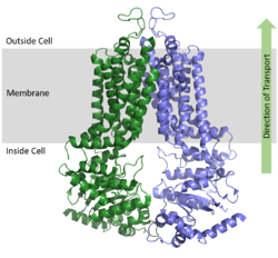
| - | ABCG2 is a homodimer with each monomer | + | ABCG2 is a homodimer with each monomer contributing two sequential regions to two overall distinct structural domains, the nucleotide binding domain <scene name='83/832932/Overall_structure_nbd_unbound/6'>(NBD)</scene> and the transmembrane domain <scene name='83/832932/Overall_structure_tmd_unbound/5'>(TMD)</scene>.<ref name="Taylor"/> The NBD binds and processes ATP and is located inside of the cell where it is exposed to the cytosol. The TMD is responsible for binding and transporting any foreign substrates and is embedded in the cell membrane and extends into the extracellular region (Figure 1). [[Image:Protein_orientation_map ABCG2.png|250 px|right|thumb|Figure 1. Orientation of ABCG2 in relation to the cell membrane. [https://www.rcsb.org/structure/5NJ3 (5NJ3)]]] |
===ATP Bound and Unbound Conformations=== | ===ATP Bound and Unbound Conformations=== | ||
| - | As an [https://en.wikipedia.org/wiki/ATP-binding_cassette_transporter ABC Transporter], ABCG2 exhibits ATPase activity by using the energy of ATP hydrolysis to facilitate transport. After substrates bind in the TMD, one molecule of <scene name='83/832932/Atp_bound_use2/3'>ATP binds each NBD</scene> (2 molecules of ATP total) causing a conformational change of the overall structure from an <scene name='83/832932/Overall_use_2/3'>inward-facing conformation</scene> to an <scene name='83/832932/Outward_facing_conformation/4'>outward-facing conformation</scene>. <scene name='83/832937/Atp_and_mg_bound_to_abcg2/4'>ATP coordinates</scene> with various residues and a magnesium ion in the <scene name='83/832932/Atp_bound_in_nbd/ | + | As an [https://en.wikipedia.org/wiki/ATP-binding_cassette_transporter ABC Transporter], ABCG2 exhibits ATPase activity by using the energy of ATP hydrolysis to facilitate transport. After substrates bind in the TMD, one molecule of <scene name='83/832932/Atp_bound_use2/3'>ATP binds each NBD</scene> (2 molecules of ATP total) causing a conformational change of the overall structure from an <scene name='83/832932/Overall_use_2/3'>inward-facing conformation</scene> to an <scene name='83/832932/Outward_facing_conformation/4'>outward-facing conformation</scene>. <scene name='83/832937/Atp_and_mg_bound_to_abcg2/4'>ATP coordinates</scene> with various residues and a magnesium ion in the <scene name='83/832932/Atp_bound_in_nbd/3'>binding site of each NBD</scene> which is bordered with [https://en.wikipedia.org/wiki/Walker_motifs Walker A and B motifs]. One molecule of ATP is hydrolyzed to transport substrates across the cell membrane while the second molecule of ATP is subsequently hydrolyzed to reset the transporter to its inward-facing conformation.<ref name="Robey"/> |
When ATP binds, α-helices in the NBD <scene name='83/832932/Atp_bound_nbd/3'>rotate</scene> approximately 35° relative to the <scene name='83/832932/Overall_structure_nbd_unbound/5'>inward-facing conformation of NBD</scene>. This shift in the NBD causes slight shifts of α-helices in the TMD; these helices are <scene name='83/832932/Atp_bound_use_tmd/4'>pushed toward each other</scene> relative to the <scene name='83/832932/Overall_structure_tmd_unbound/4'>inward-facing conformation of TMD</scene>. The overall shift from inward-facing to outward-facing promotes the transport of substrates through the transporter.<ref name="Manolaridis"/> | When ATP binds, α-helices in the NBD <scene name='83/832932/Atp_bound_nbd/3'>rotate</scene> approximately 35° relative to the <scene name='83/832932/Overall_structure_nbd_unbound/5'>inward-facing conformation of NBD</scene>. This shift in the NBD causes slight shifts of α-helices in the TMD; these helices are <scene name='83/832932/Atp_bound_use_tmd/4'>pushed toward each other</scene> relative to the <scene name='83/832932/Overall_structure_tmd_unbound/4'>inward-facing conformation of TMD</scene>. The overall shift from inward-facing to outward-facing promotes the transport of substrates through the transporter.<ref name="Manolaridis"/> | ||
| - | The NBDs in ABCG2 remain in contact with one another even without a bound substrate, providing greater substrate specificity as the entrance to the transporter is not as globular as other ABC transporters like ABCB1 or ABCC1. The entrance from the cytoplasm to the transporter is lined by [https://en.wikipedia.org/wiki/Hydrophobe hydrophobic] residues<scene name='83/832939/Lining_of_entrance_of_nbd/ | + | The NBDs in ABCG2 remain in contact with one another even without a bound substrate, providing greater substrate specificity as the entrance to the transporter is not as globular as other ABC transporters like ABCB1 or ABCC1. The entrance from the cytoplasm to the transporter is lined by [https://en.wikipedia.org/wiki/Hydrophobe hydrophobic] residues<scene name='83/832939/Lining_of_entrance_of_nbd/2'> A397, V401, L405, L539, I543, and T547</scene> in both monomers. |
===Cavities and Leucine Plug=== | ===Cavities and Leucine Plug=== | ||
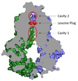
[[Image:Cavities_and_leucine_plug_abcg2.png|250 px|right|thumb|Figure 2. Locations of Cavities 1 and 2 and the Leucine Plug in ABCG2. The protein is in the inward-facing conformation with Cavity 1 open to the cytosol for substrate recruitment, the Leucine Plug is intact, and Cavity 2 is completely occluded. [https://www.rcsb.org/structure/5NJ3 (5NJ3)]]] | [[Image:Cavities_and_leucine_plug_abcg2.png|250 px|right|thumb|Figure 2. Locations of Cavities 1 and 2 and the Leucine Plug in ABCG2. The protein is in the inward-facing conformation with Cavity 1 open to the cytosol for substrate recruitment, the Leucine Plug is intact, and Cavity 2 is completely occluded. [https://www.rcsb.org/structure/5NJ3 (5NJ3)]]] | ||
| - | Substrates are transported through ABCG2 via two cavities separated by a leucine plug (Figure 2). <scene name='83/832932/Highlight_cavity_1/3'>Cavity 1</scene> acts as a multidrug binding pocket and is formed by helices at the interface of the monomers in the TMD. When ATP is not bound to the NBDs, Cavity 1 is | + | Substrates are transported through ABCG2 via two cavities separated by a leucine plug (Figure 2). <scene name='83/832932/Highlight_cavity_1/3'>Cavity 1</scene> acts as a multidrug binding pocket and is formed by helices at the interface of the monomers in the TMD. When ATP is not bound to the NBDs, Cavity 1 is open to the cytosol in order to recruit substrates for transport. Cavity 1 is <scene name='83/832932/Cavity_1_narrow_surface/4'>narrow</scene> and full of nonpolar, hydrophobic residues and, as a result, prefers nonpolar, hydrophobic substrates, particularly flat, polycyclic molecules. Substrates, such as estrone sulfate, <scene name='83/832932/Cavity_1_-_use2/4'>form hydrogen bonds and stacking interactions</scene> with residues from each subunit in Cavity 1.<ref name="Taylor"/> |
| - | After substrates bind in Cavity 1, ATP binds each NBD leading to the transporter shifting from inward-facing to outward-facing. The outward-facing conformation results in the <scene name='83/832932/Atp_bound_cavity_2/3'>collapse of Cavity 1</scene> in the TMD in which the cavity is no longer <scene name='83/832932/Overall_structure_cavity_1hel/4'>open to the cytosol</scene>. This collapse forces the substrate | + | After substrates bind in Cavity 1, ATP binds each NBD leading to the transporter shifting from inward-facing to outward-facing. The outward-facing conformation results in the <scene name='83/832932/Atp_bound_cavity_2/3'>collapse of Cavity 1</scene> in the TMD in which the cavity is no longer <scene name='83/832932/Overall_structure_cavity_1hel/4'>open to the cytosol</scene>. This collapse forces the substrate into Cavity 2 as there is no longer room in Cavity 1 to accommodate substrates.<ref name="Manolaridis"/> <scene name='83/832932/Atp_bound_use_cav_2/3'>Cavity 2</scene>, which is occluded when the protein in is the inward-facing conformation, is now open to the extracellular space and able to release the substrate. Cavity 2 contains a less hydrophobic environment and, as a result, substrates are released due to hydrophobic mismatch.<ref name="Taylor"/> <scene name='83/832932/Atp_bound_use_el_disulfides/3'>Disulfide bonds</scene> in the external loops near the exit of Cavity 2 also help promote substrate release.<ref name="Manolaridis"/> Once Cavity 2 is empty, the protein reverts back to the inward-facing conformation via hydrolysis of ATP. |
| - | Cavities 1 and 2 are separated by a <scene name='83/832932/Leucine_plug_open_con/7'>leucine plug</scene> which likely acts as a substrate check-point during transport | + | Cavities 1 and 2 are separated by a <scene name='83/832932/Leucine_plug_open_con/7'>leucine plug</scene> which likely acts as a substrate check-point during transport. Changes to either of these leucine residues have exhibited an increase in transport and a decrease in substrate specificity.<ref name="Manolaridis"/> After the substrate binds Cavity 1 and ATP molecules bind each NBD, the <scene name='83/832932/Atp_bound_cavsleu/5'>leucine plug opens</scene> to allow the substrate to enter Cavity 2. Once the substrate enters Cavity 2, the plug is able to reform and promote substrate release and conversion to the inward-facing conformation. |
===Structural Elucidation of ABCG2=== | ===Structural Elucidation of ABCG2=== | ||
| - | Binding of two antigen binding fragments (<scene name='83/832939/Abcg2_with_bound_5d3-fab/4'>5D3-Fab</scene>) were required for stabilization of ABCG2 for high resolution cryo-EM images to be developed for the ABCG2 transporter protein.<ref name="Taylor"/><ref name="Manolaridis"/> 5D3 Fab <scene name='83/832939/Fab_binding_site/ | + | Binding of two [https://en.wikipedia.org/wiki/Fragment_antigen-binding antigen binding fragments](<scene name='83/832939/Abcg2_with_bound_5d3-fab/4'>5D3-Fab</scene>) were required for stabilization of ABCG2 for high resolution cryo-EM images to be developed for the ABCG2 transporter protein.<ref name="Taylor"/><ref name="Manolaridis"/> 5D3 Fab <scene name='83/832939/Fab_binding_site/3'>clamps</scene> the two domains together preventing movement of the transporter from inward to outward facing. Fab binds at a 35 degree angle relative to the membrane plane which stops the 40 degree transition of the TMD from closed to open. Interestingly, complete arrest of ABCG2 in its inward facing state only requires one Fab bound. However in stabilization experiments, two Fabs were found to be bound to each monomer.<ref name="Taylor"/><ref name="Manolaridis"/> |
==Disease== | ==Disease== | ||
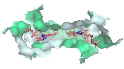
[[Image:Ligand_Interactions_6ffc.png|400 px|right|thumb|Figure 3: MZ29 bound to cavity 1 of ABCG2 [https://www.rcsb.org/structure/6FFC (6FFC)]. Two MZ29 are shown in sticks and are colored by element. Hydrophobic interactions between the surface of cavity 1 and MZ29 are shown in green.]] | [[Image:Ligand_Interactions_6ffc.png|400 px|right|thumb|Figure 3: MZ29 bound to cavity 1 of ABCG2 [https://www.rcsb.org/structure/6FFC (6FFC)]. Two MZ29 are shown in sticks and are colored by element. Hydrophobic interactions between the surface of cavity 1 and MZ29 are shown in green.]] | ||
| - | Dysfunctions in ABCG2 are linked to [https://en.wikipedia.org/wiki/Hyperuricemia hyperuricemia] which can lead to [https://en.wikipedia.org/wiki/Gout gout], [https://en.wikipedia.org/wiki/Kidney_disease kidney disease], and [https://en.wikipedia.org/wiki/Hypertension hypertension], all of which are thought to be the result of impaired transport of uric acid. Additionally, the expression of ABCG2 has been found to correlate with a poor prognosis and treatment outcome of various cancers including breast, ovarian, and lung.<ref name="Jackson"/> | + | Dysfunctions in ABCG2 are linked to [https://en.wikipedia.org/wiki/Hyperuricemia hyperuricemia] which can lead to [https://en.wikipedia.org/wiki/Gout gout], [https://en.wikipedia.org/wiki/Kidney_disease kidney disease], and [https://en.wikipedia.org/wiki/Hypertension hypertension], all of which are thought to be the result of impaired transport of uric acid. Additionally, the expression of ABCG2 has been found to correlate with a poor prognosis and treatment outcome of various cancers including breast, ovarian, and lung.<ref name="Jackson">PMID:29610494</ref> |
| - | Several mutations also decrease transporter activity.<ref name="Taylor"/><ref name="Manolaridis"/><ref name="Robey"/> The most detrimental of these is a point mutation of <scene name='83/832939/Residue_211/1'> | + | Several mutations also decrease transporter activity.<ref name="Taylor"/><ref name="Manolaridis"/><ref name="Robey"/> The most detrimental of these is a point mutation of <scene name='83/832939/Residue_211/1'>Glu211 to Gln211</scene> , which completely abolished activity of the transporter. Another point mutation occurs <scene name='83/832939/The_cause_of_gout/1'>at Gln141</scene>, which when mutated to lysine causes gout by distorting ABCG2's tertiary structure. Changes at <scene name='83/832939/R482/1'>Arg482</scene> shift the substrate specificity of ABCG2 by [https://en.wikipedia.org/wiki/Allosteric_regulation allosteric effects] as this residue is distantly located from the binding pocket. |
===Cancer=== | ===Cancer=== | ||
| - | ABCG2 hinders cancer | + | ABCG2 hinders cancer inhibition by contributing to [https://en.wikipedia.org/wiki/Multiple_drug_resistance multidrug resistance] in tumor cells. ABCG2 exports xenbiotics, including vital anti-cancer drugs, which results in the inability to treat cancer cells. Cancer patients often show high levels of expression of multiple ABC transporters. For example, [https://en.wikipedia.org/wiki/Acute_myeloid_leukemia acute myeloid leukemia] (AML) has an increased expression of [https://en.wikipedia.org/wiki/P-glycoprotein ABCB1], [https://en.wikipedia.org/wiki/ABCG1 ABCG1], and ABCG2 while childhood AML shows an increased expression in [https://en.wikipedia.org/wiki/ABCA3 ABCA3], ABCB1, [https://en.wikipedia.org/wiki/ABCC3 ABCC3], and ABCG2.<ref name="Marzac">PMID:21606172</ref><ref name="Bartholomae">PMID:26512967</ref> Additionally, pancreatic cancer has shown an upregulation of [https://en.wikipedia.org/wiki/ABCB4 ABCB4], [https://en.wikipedia.org/wiki/ABCB11 ABCB11], [https://en.wikipedia.org/wiki/ABCC1 ABCC1], ABCC3, [https://en.wikipedia.org/wiki/ABCC5 ABCC5], [https://en.wikipedia.org/wiki/ABCC10 ABCC10], and ABCG2.<ref name="Mohelnikova-Duchonova">PMID:23462326</ref> |
The substrate specificity among ABC transporters varies, so this protein family can collectively export a wide variety of substrates and, ultimately, a wide variety of anticancer drugs (Figures 3 and 4). ABCG2 has been known to export anticancer drugs such [https://en.wikipedia.org/wiki/Methotrexate methotrexate], [https://en.wikipedia.org/wiki/Mitoxantrone mitoxantrone], [https://en.wikipedia.org/wiki/Topotecan topotecan], [https://en.wikipedia.org/wiki/Irinotecan irinotecan], and [https://en.wikipedia.org/wiki/Alvocidib flavopiridol]<ref name="Mao">PMID:25236865</ref>. Due to the high expression of multiple ABC transporters in cancer cells, simultaneous treatment of multiple transporters would likely be necessary for successful cancer treatment. | The substrate specificity among ABC transporters varies, so this protein family can collectively export a wide variety of substrates and, ultimately, a wide variety of anticancer drugs (Figures 3 and 4). ABCG2 has been known to export anticancer drugs such [https://en.wikipedia.org/wiki/Methotrexate methotrexate], [https://en.wikipedia.org/wiki/Mitoxantrone mitoxantrone], [https://en.wikipedia.org/wiki/Topotecan topotecan], [https://en.wikipedia.org/wiki/Irinotecan irinotecan], and [https://en.wikipedia.org/wiki/Alvocidib flavopiridol]<ref name="Mao">PMID:25236865</ref>. Due to the high expression of multiple ABC transporters in cancer cells, simultaneous treatment of multiple transporters would likely be necessary for successful cancer treatment. | ||
| Line 47: | Line 47: | ||
Due to the potential for ABCG2 inhibition to aid in cancer treatment, efforts have been made to develop specific inhibitors of ABCG2 and other ABC transporters. The ABC transporter ABCB1, also known as multidrug resistance 1 (MDR1), was a therapeutic target in previous studies which produced three generations of MDR1 inhibitors, such as [https://en.wikipedia.org/wiki/Verapamil verapamil], [https://en.wikipedia.org/wiki/Valspodar valspodar], and [https://en.wikipedia.org/wiki/Zosuquidar zosuquidar]; however, many of these inhibitors had neurotoxic effects that discouraged their use in cancer treatment.<ref name="Leonard">PMID:14530494</ref><ref name="Binkhathlan">PMID:23369096</ref><ref name="Witherspoon">PMID:9816083</ref> | Due to the potential for ABCG2 inhibition to aid in cancer treatment, efforts have been made to develop specific inhibitors of ABCG2 and other ABC transporters. The ABC transporter ABCB1, also known as multidrug resistance 1 (MDR1), was a therapeutic target in previous studies which produced three generations of MDR1 inhibitors, such as [https://en.wikipedia.org/wiki/Verapamil verapamil], [https://en.wikipedia.org/wiki/Valspodar valspodar], and [https://en.wikipedia.org/wiki/Zosuquidar zosuquidar]; however, many of these inhibitors had neurotoxic effects that discouraged their use in cancer treatment.<ref name="Leonard">PMID:14530494</ref><ref name="Binkhathlan">PMID:23369096</ref><ref name="Witherspoon">PMID:9816083</ref> | ||
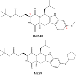
| - | While ABC transporter inhibition was dismissed after failed clinical trials, interest in revisiting ABC inhibition has reemerged due to new developments made in recent years.<ref name="Robey"/> For instance, Kol143 (Figure 4) is a compound derived from fungal toxin [https://en.wikipedia.org/wiki/Fumitremorgin fumitremorgin C] (FTC), a selective inhibitor of ABCG2 which exhibits undesirable neurotoxic effects.<ref name="Allen">PMID:12477054</ref> Kol143 was found to be less toxic and more potent than FTC; however, this inhibitor is nonselective toward ABCG2.<ref name="Weidner">PMID:26148857</ref> Various inhibitors were derived from Kol143 with changes made at positions 1 and 2 in Figure | + | While ABC transporter inhibition was dismissed after failed clinical trials, interest in revisiting ABC inhibition has reemerged due to new developments made in recent years.<ref name="Robey"/> For instance, Kol143 (Figure 4) is a compound derived from fungal toxin [https://en.wikipedia.org/wiki/Fumitremorgin fumitremorgin C] (FTC), a selective inhibitor of ABCG2 which exhibits undesirable neurotoxic effects.<ref name="Allen">PMID:12477054</ref> Kol143 was found to be less toxic and more potent than FTC; however, this inhibitor is nonselective toward ABCG2.<ref name="Weidner">PMID:26148857</ref> Various inhibitors were derived from Kol143 with changes made at positions 1 and 2 indicated in Figure 4. Modifications at these positions prove to affect the inhibitory capacities of compounds. A promising compound which has shown a high degree of potency is the inhibitor MZ29 (Figures 3 and 4).<ref name="Jackson"/> |
| - | ABCG2 inhibitors, such as <scene name='83/832932/Inhibitor_bound_cavity_1/2'>MZ29</scene> | + | ABCG2 inhibitors, such as <scene name='83/832932/Inhibitor_bound_cavity_1/2'>MZ29</scene> (Figure 3) bind in Cavity 1. They act as competitive inhibitors against ABCG2 substrates and show a higher affinity toward the transporter. Depending on the size of the inhibitor, one or two molecules can accommodate binding to the cavity and form <scene name='83/832932/Inhibitor_interactions_cavity1/3'>hydrogen bonds, van der Waals, and stacking interactions</scene> within the binding site.<ref name="Jackson"/> Many inhibitors are too big to be transported past the leucine plug resulting in the "clogging" of the transporter. With inhibitors acting as wedges, ABCG2 is locked in the inward-facing conformation and unable to transport xenobiotics out of the cell.<ref name="Manolaridis"/> |
</StructureSection> | </StructureSection> | ||
== References == | == References == | ||
Current revision
ABCG2 Multidrug Transporter
References
- ↑ 1.0 1.1 1.2 1.3 1.4 1.5 1.6 Taylor NMI, Manolaridis I, Jackson SM, Kowal J, Stahlberg H, Locher KP. Structure of the human multidrug transporter ABCG2. Nature. 2017 Jun 22;546(7659):504-509. doi: 10.1038/nature22345. Epub 2017 May, 29. PMID:28554189 doi:http://dx.doi.org/10.1038/nature22345
- ↑ 2.0 2.1 2.2 2.3 2.4 2.5 2.6 2.7 2.8 Manolaridis I, Jackson SM, Taylor NMI, Kowal J, Stahlberg H, Locher KP. Cryo-EM structures of a human ABCG2 mutant trapped in ATP-bound and substrate-bound states. Nature. 2018 Nov;563(7731):426-430. doi: 10.1038/s41586-018-0680-3. Epub 2018 Nov, 7. PMID:30405239 doi:http://dx.doi.org/10.1038/s41586-018-0680-3
- ↑ 3.0 3.1 3.2 3.3 Robey RW, Pluchino KM, Hall MD, Fojo AT, Bates SE, Gottesman MM. Revisiting the role of ABC transporters in multidrug-resistant cancer. Nat Rev Cancer. 2018 Jul;18(7):452-464. doi: 10.1038/s41568-018-0005-8. PMID:29643473 doi:http://dx.doi.org/10.1038/s41568-018-0005-8
- ↑ 4.0 4.1 4.2 Jackson SM, Manolaridis I, Kowal J, Zechner M, Taylor NMI, Bause M, Bauer S, Bartholomaeus R, Bernhardt G, Koenig B, Buschauer A, Stahlberg H, Altmann KH, Locher KP. Structural basis of small-molecule inhibition of human multidrug transporter ABCG2. Nat Struct Mol Biol. 2018 Apr;25(4):333-340. doi: 10.1038/s41594-018-0049-1. Epub, 2018 Apr 2. PMID:29610494 doi:http://dx.doi.org/10.1038/s41594-018-0049-1
- ↑ Marzac C, Garrido E, Tang R, Fava F, Hirsch P, De Benedictis C, Corre E, Lapusan S, Lallemand JY, Marie JP, Jacquet E, Legrand O. ATP Binding Cassette transporters associated with chemoresistance: transcriptional profiling in extreme cohorts and their prognostic impact in a cohort of 281 acute myeloid leukemia patients. Haematologica. 2011 Sep;96(9):1293-301. doi: 10.3324/haematol.2010.031823. Epub, 2011 May 23. PMID:21606172 doi:http://dx.doi.org/10.3324/haematol.2010.031823
- ↑ Bartholomae S, Gruhn B, Debatin KM, Zimmermann M, Creutzig U, Reinhardt D, Steinbach D. Coexpression of Multiple ABC-Transporters is Strongly Associated with Treatment Response in Childhood Acute Myeloid Leukemia. Pediatr Blood Cancer. 2016 Feb;63(2):242-7. doi: 10.1002/pbc.25785. Epub 2015 Oct, 29. PMID:26512967 doi:http://dx.doi.org/10.1002/pbc.25785
- ↑ Mohelnikova-Duchonova B, Brynychova V, Oliverius M, Honsova E, Kala Z, Muckova K, Soucek P. Differences in transcript levels of ABC transporters between pancreatic adenocarcinoma and nonneoplastic tissues. Pancreas. 2013 May;42(4):707-16. doi: 10.1097/MPA.0b013e318279b861. PMID:23462326 doi:http://dx.doi.org/10.1097/MPA.0b013e318279b861
- ↑ Mao Q, Unadkat JD. Role of the breast cancer resistance protein (BCRP/ABCG2) in drug transport--an update. AAPS J. 2015 Jan;17(1):65-82. doi: 10.1208/s12248-014-9668-6. Epub 2014 Sep 19. PMID:25236865 doi:http://dx.doi.org/10.1208/s12248-014-9668-6
- ↑ Leonard GD, Fojo T, Bates SE. The role of ABC transporters in clinical practice. Oncologist. 2003;8(5):411-24. doi: 10.1634/theoncologist.8-5-411. PMID:14530494 doi:http://dx.doi.org/10.1634/theoncologist.8-5-411
- ↑ Binkhathlan Z, Lavasanifar A. P-glycoprotein inhibition as a therapeutic approach for overcoming multidrug resistance in cancer: current status and future perspectives. Curr Cancer Drug Targets. 2013 Mar;13(3):326-46. doi:, 10.2174/15680096113139990076. PMID:23369096 doi:http://dx.doi.org/10.2174/15680096113139990076
- ↑ Witherspoon SM, Emerson DL, Kerr BM, Lloyd TL, Dalton WS, Wissel PS. Flow cytometric assay of modulation of P-glycoprotein function in whole blood by the multidrug resistance inhibitor GG918. Clin Cancer Res. 1996 Jan;2(1):7-12. PMID:9816083
- ↑ Allen JD, van Loevezijn A, Lakhai JM, van der Valk M, van Tellingen O, Reid G, Schellens JH, Koomen GJ, Schinkel AH. Potent and specific inhibition of the breast cancer resistance protein multidrug transporter in vitro and in mouse intestine by a novel analogue of fumitremorgin C. Mol Cancer Ther. 2002 Apr;1(6):417-25. PMID:12477054
- ↑ Weidner LD, Zoghbi SS, Lu S, Shukla S, Ambudkar SV, Pike VW, Mulder J, Gottesman MM, Innis RB, Hall MD. The Inhibitor Ko143 Is Not Specific for ABCG2. J Pharmacol Exp Ther. 2015 Sep;354(3):384-93. doi: 10.1124/jpet.115.225482. Epub , 2015 Jul 6. PMID:26148857 doi:http://dx.doi.org/10.1124/jpet.115.225482
Student Contributors
Julia Pomeroy
Shelby Skaggs
Sam Sullivan
Jaelyn Voyles


