Journal:Acta Cryst D:S205979832000772X
From Proteopedia
(Difference between revisions)

| (31 intermediate revisions not shown.) | |||
| Line 1: | Line 1: | ||
| - | <StructureSection load='' size='450' side='right' scene=' | + | <StructureSection load='' size='450' side='right' scene='85/853345/Cv/1' caption=''> |
===Structural evidence for mono- and di-carboxylates binding at pharmacologically relevant extracellular sites of a pentameric ligand gated ion channel=== | ===Structural evidence for mono- and di-carboxylates binding at pharmacologically relevant extracellular sites of a pentameric ligand gated ion channel=== | ||
<big>Zaineb Fourati, Ludovic Saugueta, and Marc Delarue</big> <ref>doi 10.1107/S205979832000772X</ref> | <big>Zaineb Fourati, Ludovic Saugueta, and Marc Delarue</big> <ref>doi 10.1107/S205979832000772X</ref> | ||
<hr/> | <hr/> | ||
<b>Molecular Tour</b><br> | <b>Molecular Tour</b><br> | ||
| + | Pentameric ligand-gated ion channels (pLGICs) are key receptors mediating electrochemical signaling in the nervous system. These receptors, that include the well-known GABAA and acetylcholine receptors, are activated by agonist/neurotransmitter binding to a specific orthosteric site in the extracellular domain, leading to channel opening and ion permeation inside the cell. They are organised as pentamers of identical or homologous subunits that respond in a concerted manner to agonist binding in the orthosteric site. Beyond the activation process, these receptors also respond to various chemical stimuli such as alcohol as well as some drugs that modulate the receptor function. Because these molecules bind and act at a distinct site from the orthosteric agonist site, they are classified as allosteric modulators. This allosteric modulation is a key feature of pLGIC pharmacological response. A comprehensive characterization of the allosteric modulation sites within pLGICs is thus a prerequisite toward the rational conception of efficient drugs targeting these receptors. In this study, we identify and characterize two modulation sites in the extracellular domain of GLIC, a bacterial pLGIC exhibiting similar pharmacological properties as its eukaryotic counterparts, especially cationic ones. In particular, we show that these pockets can accommodate different mono- and di- acetic acid derivatives, some of which are known to be allosteric inhibitors of GLIC. One of them, called the intersubunit site, partially overlaps the orthosteric (agonist) site; the other one is located behind the first one, is facing the interior of the vestibule and is called intrasubunit site. | ||
| + | |||
| + | The X-ray structure of GLIC in complex with succinate (PDB code [[6hjz]]): | ||
| + | *<scene name='85/853345/Cv/5'>Receptor side view</scene>. | ||
| + | *<scene name='85/853345/Cv/6'>Receptor top view</scene>. The GLIC receptor is represented with white traces. Succinate binds 5 intrasubunit (cyan spacefill) and 5 intersubunit (hotpink spacefill) sites in the extracellular domain of GLIC. | ||
| + | *<scene name='85/853345/Cv/8'>Receptor side view. Subunits are colored in different colors</scene>. | ||
| + | *<scene name='85/853345/Cv/10'>Receptor top view. Subunits are colored in different colors</scene>. | ||
| + | *<scene name='85/853345/Cv/14'>Succinate binds in intrasubunit site only with residues from one subunit</scene>. | ||
| + | *<scene name='85/853345/Cv/15'>Succinate binds in intersubunit site with residues from 2 adjacent subunits</scene>. | ||
| + | |||
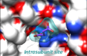
| + | These extracellular sites are of great interest because they can accommodate drugs distinct from the common hydrophobic general anaesthetics that find their binding site through diffusion in the plasma membrane. Moreover, one of these sites, namely the intrasubunit one, is conserved in other cationic pLGICs such as the serotonin receptor, allowing a more specific targeting of cationic receptors. <scene name='85/853345/Cv/24'>Mapping the intrasubunit succinate binding-site to the X-ray structure of the 5HT3 serotonin receptor</scene> (PDB code [[4pir]]). See also the static image below: | ||
| + | [[Image:Figel2.png|thumb|left|Succinate is shown as purple sticks]] | ||
| + | {{Clear}} | ||
| + | This latter site is likely to be a key pharmacological site and thus an important target for rational drug design against some neurological disorders involving human pLGICs. | ||
| + | |||
| + | '''PDB references:''' Xray structure of GLIC in complex with fumarate [[6hj3]]; Xray structure of GLIC in complex with glutarate [[6hja]]; Xray structure of GLIC in complex with malonate [[6hjb]]; Xray structure of GLIC in complex with crotonate [[6hji]]; Xray structure of GLIC in complex with succinate [[6hjz]]; X-ray structure of GLIC in complex with propionate [[6hpp]]. | ||
<b>References</b><br> | <b>References</b><br> | ||
Current revision
| |||||||||||
This page complements a publication in scientific journals and is one of the Proteopedia's Interactive 3D Complement pages. For aditional details please see I3DC.

