Mitochondrial calcium uniporter
From Proteopedia
(Difference between revisions)
(New page: ==Mitochondrial Calcium Uniporter== <StructureSection load='6dnf' size='340' side='right' caption='Mitochondrial Calcium Uniporter (MCU): Each monomer is shown in a different color. Calciu...) |
(Category:Featured in BAMBED) |
||
| (6 intermediate revisions not shown.) | |||
| Line 1: | Line 1: | ||
| + | {{BAMBED | ||
| + | |DATE=December 3, 2020 | ||
| + | |OLDID=3326279 | ||
| + | |BAMBEDDOI=10.1002/bmb.21560 | ||
| + | }} | ||
| + | |||
==Mitochondrial Calcium Uniporter== | ==Mitochondrial Calcium Uniporter== | ||
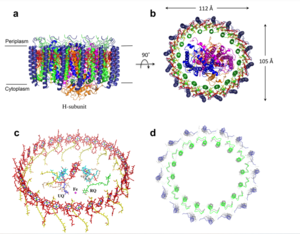
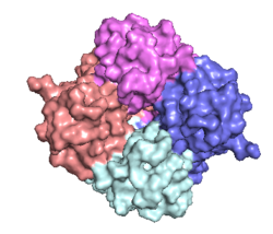
<StructureSection load='6dnf' size='340' side='right' caption='Mitochondrial Calcium Uniporter (MCU): Each monomer is shown in a different color. Calcium ions are shown in green. (PDB Code [http://www.rcsb.org/pdb/explore/explore.do?structureId=6DNF 6DNF])' scene='83/832952/Starting_scene/5'> | <StructureSection load='6dnf' size='340' side='right' caption='Mitochondrial Calcium Uniporter (MCU): Each monomer is shown in a different color. Calcium ions are shown in green. (PDB Code [http://www.rcsb.org/pdb/explore/explore.do?structureId=6DNF 6DNF])' scene='83/832952/Starting_scene/5'> | ||
== Overview == | == Overview == | ||
| - | The mitochondrial calcium uniporter (MCU) complex is the main source of entry for [https://en.wikipedia.org/wiki/Calcium calcium] ions into the [https://en.wikipedia.org/wiki/Mitochondrial_matrix mitochondrial matrix] from the [https://en.wikipedia.org/wiki/Mitochondrion#Intermembrane_space intermembrane space]. MCU channels exist in most [https://en.wikipedia.org/wiki/Eukaryote eukaryotes], but activity is regulated differently in each [https://en.wikipedia.org/wiki/Clade clade].<ref name="Baradaran">PMID:29995857</ref> MCU was definitively assigned in 2011 using a combination of [https://en.wikipedia.org/wiki/Nuclear_magnetic_resonance_spectroscopy NMR spectroscopy], [https://en.wikipedia.org/wiki/Cryogenic_electron_microscopy cryoelectron microscopy], and [https://en.wikipedia.org/wiki/X-ray_crystallography x-ray crystallography].<ref name="Woods">PMID:31869674</ref> Recent [https://en.wikipedia.org/wiki/Cryogenic_electron_microscopy cryoelectron microscopy] (cryo-EM) analysis provides a structural framework for understanding the mechanism for calcium selectivity by the MCU.<ref name="Giorgi" /> Like other ion channels, the MCU is highly selective and efficient, allowing calcium ions into the mitochondrial matrix at a rate of 5,000,000 ions per second, even though [https://en.wikipedia.org/wiki/Potassium potassium] ions are over 100,000 times more abundant in the intermembrane space.<ref name="Baradaran"/> | + | The '''mitochondrial calcium uniporter''' (MCU) complex is the main source of entry for [https://en.wikipedia.org/wiki/Calcium calcium] ions into the [https://en.wikipedia.org/wiki/Mitochondrial_matrix mitochondrial matrix] from the [https://en.wikipedia.org/wiki/Mitochondrion#Intermembrane_space intermembrane space]. MCU channels exist in most [https://en.wikipedia.org/wiki/Eukaryote eukaryotes], but activity is regulated differently in each [https://en.wikipedia.org/wiki/Clade clade].<ref name="Baradaran">PMID:29995857</ref> MCU was definitively assigned in 2011 using a combination of [https://en.wikipedia.org/wiki/Nuclear_magnetic_resonance_spectroscopy NMR spectroscopy], [https://en.wikipedia.org/wiki/Cryogenic_electron_microscopy cryoelectron microscopy], and [https://en.wikipedia.org/wiki/X-ray_crystallography x-ray crystallography].<ref name="Woods">PMID:31869674</ref> Recent [https://en.wikipedia.org/wiki/Cryogenic_electron_microscopy cryoelectron microscopy] (cryo-EM) analysis provides a structural framework for understanding the mechanism for calcium selectivity by the MCU.<ref name="Giorgi" /> Like other ion channels, the MCU is highly selective and efficient, allowing calcium ions into the mitochondrial matrix at a rate of 5,000,000 ions per second, even though [https://en.wikipedia.org/wiki/Potassium potassium] ions are over 100,000 times more abundant in the intermembrane space.<ref name="Baradaran"/> |
Under resting conditions, the calcium concentration in the mitochondria is about the same as in the [https://en.wikipedia.org/wiki/Cytoplasm cytoplasm], but when stimulated, mitochondrial calcium concentration increases 10 to 20-fold.<ref name="Giorgi">PMID:30143745</ref> [https://en.wikipedia.org/wiki/Mitochondria_associated_membranes Mitochondria-associated ER membranes] exist between the mitochondria and the [https://en.wikipedia.org/wiki/Endoplasmic_reticulum endoplasmic reticulum] facilitate efficient transport of calcium ions.<ref name="Wang">PMID:28882140</ref> The transfer of electrons through [https://en.wikipedia.org/wiki/Electron_transport_chain#Mitochondrial_redox_carriers respiratory complexes I-IV] produces the energy to pump [https://en.wikipedia.org/wiki/Hydrogen_ion hydrogen ions] into the intermembrane space and establish the proton [https://en.wikipedia.org/wiki/Electrochemical_gradient electrochemical gradient] potential.<ref name="Giorgi"/> This negative electrochemical potential is the driving force that moves positively charged calcium ions into the mitochondrial matrix.<ref name="Giorgi"/> Calcium uptake and efflux must be tightly regulated to controll essential [https://en.wikipedia.org/wiki/Citric_acid_cycle Krebs cycle] enzyme activity, including [http://proteopedia.org/wiki/index.php/Pyruvate_dehydrogenase pyruvate dehydrogenase], [https://en.wikipedia.org/wiki/Oxoglutarate_dehydrogenase_complex α-ketoglutarate dehydrogenase], and [http://proteopedia.org/wiki/index.php/Isocitrate_dehydrogenase isocitrate dehydrogenase], while avoiding calcium overload and [https://en.wikipedia.org/wiki/Apoptosis apoptosis].<ref name="Wang"/> | Under resting conditions, the calcium concentration in the mitochondria is about the same as in the [https://en.wikipedia.org/wiki/Cytoplasm cytoplasm], but when stimulated, mitochondrial calcium concentration increases 10 to 20-fold.<ref name="Giorgi">PMID:30143745</ref> [https://en.wikipedia.org/wiki/Mitochondria_associated_membranes Mitochondria-associated ER membranes] exist between the mitochondria and the [https://en.wikipedia.org/wiki/Endoplasmic_reticulum endoplasmic reticulum] facilitate efficient transport of calcium ions.<ref name="Wang">PMID:28882140</ref> The transfer of electrons through [https://en.wikipedia.org/wiki/Electron_transport_chain#Mitochondrial_redox_carriers respiratory complexes I-IV] produces the energy to pump [https://en.wikipedia.org/wiki/Hydrogen_ion hydrogen ions] into the intermembrane space and establish the proton [https://en.wikipedia.org/wiki/Electrochemical_gradient electrochemical gradient] potential.<ref name="Giorgi"/> This negative electrochemical potential is the driving force that moves positively charged calcium ions into the mitochondrial matrix.<ref name="Giorgi"/> Calcium uptake and efflux must be tightly regulated to controll essential [https://en.wikipedia.org/wiki/Citric_acid_cycle Krebs cycle] enzyme activity, including [http://proteopedia.org/wiki/index.php/Pyruvate_dehydrogenase pyruvate dehydrogenase], [https://en.wikipedia.org/wiki/Oxoglutarate_dehydrogenase_complex α-ketoglutarate dehydrogenase], and [http://proteopedia.org/wiki/index.php/Isocitrate_dehydrogenase isocitrate dehydrogenase], while avoiding calcium overload and [https://en.wikipedia.org/wiki/Apoptosis apoptosis].<ref name="Wang"/> | ||
| Line 57: | Line 63: | ||
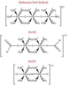
Calcium overload in the mitochondria of cardiac cells lead to [https://en.wikipedia.org/wiki/Apoptosis apoptotic] cardiac cell death. Calcium governs [https://en.wikipedia.org/wiki/Cardiac_excitation-contraction_coupling excitation contraction coupling] of the cardiac muscles, which creates the ATP needed to power the contraction during heart beats. The increase in mitochondrial Ca<sup>2+</sup> concentration is essential for the functioning of this muscle contraction. Mitochondrial Ca<sup>2+</sup> overload, though, leads to [https://en.wikipedia.org/wiki/Necrosis necrotic] cardiac cell death and can be targeted with regulation of the MCU. An example of potential treatment might involve the use of Ru360 to inhibit the uptake of Ca<sup>2+</sup> ions into the mitochondria.<ref name="Giorgi" /> | Calcium overload in the mitochondria of cardiac cells lead to [https://en.wikipedia.org/wiki/Apoptosis apoptotic] cardiac cell death. Calcium governs [https://en.wikipedia.org/wiki/Cardiac_excitation-contraction_coupling excitation contraction coupling] of the cardiac muscles, which creates the ATP needed to power the contraction during heart beats. The increase in mitochondrial Ca<sup>2+</sup> concentration is essential for the functioning of this muscle contraction. Mitochondrial Ca<sup>2+</sup> overload, though, leads to [https://en.wikipedia.org/wiki/Necrosis necrotic] cardiac cell death and can be targeted with regulation of the MCU. An example of potential treatment might involve the use of Ru360 to inhibit the uptake of Ca<sup>2+</sup> ions into the mitochondria.<ref name="Giorgi" /> | ||
</StructureSection> | </StructureSection> | ||
| + | |||
| + | ==3D structures of mitochondrial calcium uniporter== | ||
| + | |||
| + | Updated on {{REVISIONDAY2}}-{{MONTHNAME|{{REVISIONMONTH}}}}-{{REVISIONYEAR}} | ||
| + | {{#tree:id=OrganizedByTopic|openlevels=0| | ||
| + | |||
| + | *Mitochondrial calcium uniporter | ||
| + | |||
| + | **[[4xtb]] – hMCU N-terminal – human <br /> | ||
| + | **[[4xsj]], [[5bz6]] – hMCU N-terminal/T4 lysozyme<br /> | ||
| + | **[[6jg0]], [[6kvx]] – hMCU N-terminal (mutant)/T4 lysozyme<br /> | ||
| + | **[[5kug]], [[5kui]], [[5kuj]] – hMCU residues 72-189<br /> | ||
| + | **[[5kue]] – hMCU residues 72-189 (mutant)<br /> | ||
| + | **[[6eaz]] – MCU - mouse<br /> | ||
| + | **[[5id3]] – MCU pore-forming domain – ''Caenorhabditis elegans'' - NMR <br /> | ||
| + | **[[6dnf]] – MCU – ''Cyphellophora europaea'' – Cryo EM<br /> | ||
| + | **[[6dt0]] – MCU – ''Neurospora crassa'' – Cryo EM<br /> | ||
| + | **[[6d7w]] – AfMCU – ''Aspergillus fischeri'' – Cryo EM<br /> | ||
| + | **[[6x4s]] – MCU/essential MCU regulator – ''Tribolium castaneum'' – Cryo EM<br /> | ||
| + | **[[5z2h]], [[5z2i]] – MCU N-terminal – ''Dictyostelium discoideum''<br /> | ||
| + | **[[6c5r]] – MaMCU soluble domain 99-426 – ''Metarhizium acridum'' <br /> | ||
| + | |||
| + | *Mitochondrial calcium uniporter complex | ||
| + | |||
| + | **[[6k7x]], [[6o58]], [[6o5b]] – hMCU + essential MCU regulator – Cryo EM<br /> | ||
| + | **[[6wdn]], [[6wdo]], [[6k7y]], [[6xjv]], [[6xjx]], [[6xqn]] – hMCU + essential MCU regulator + calcium uptake proteins 1,2 – Cryo EM<br /> | ||
| + | **[[6d80]] – AfMCU + saposin – Cryo EM<br /> | ||
| + | **[[6c5w]] – MaMCU soluble domain + nanobody<br /> | ||
| + | }} | ||
==References== | ==References== | ||
<references/> | <references/> | ||
| Line 70: | Line 105: | ||
Rieser Wells | Rieser Wells | ||
| + | [[Category:Topic Page]] | ||
| + | [[Category:Featured in BAMBED]] | ||
Current revision
This page, as it appeared on December 3, 2020, was featured in this article in the journal Biochemistry and Molecular Biology Education.
Contents |
Mitochondrial Calcium Uniporter
| |||||||||||
3D structures of mitochondrial calcium uniporter
Updated on 28-August-2025
References
- ↑ 1.00 1.01 1.02 1.03 1.04 1.05 1.06 1.07 1.08 1.09 1.10 1.11 1.12 1.13 1.14 1.15 1.16 1.17 1.18 1.19 1.20 1.21 1.22 Baradaran R, Wang C, Siliciano AF, Long SB. Cryo-EM structures of fungal and metazoan mitochondrial calcium uniporters. Nature. 2018 Jul 11. pii: 10.1038/s41586-018-0331-8. doi:, 10.1038/s41586-018-0331-8. PMID:29995857 doi:http://dx.doi.org/10.1038/s41586-018-0331-8
- ↑ 2.0 2.1 2.2 2.3 2.4 2.5 2.6 2.7 2.8 2.9 Woods JJ, Wilson JJ. Inhibitors of the mitochondrial calcium uniporter for the treatment of disease. Curr Opin Chem Biol. 2019 Dec 20;55:9-18. doi: 10.1016/j.cbpa.2019.11.006. PMID:31869674 doi:http://dx.doi.org/10.1016/j.cbpa.2019.11.006
- ↑ 3.0 3.1 3.2 3.3 3.4 3.5 3.6 3.7 3.8 Giorgi C, Marchi S, Pinton P. The machineries, regulation and cellular functions of mitochondrial calcium. Nat Rev Mol Cell Biol. 2018 Nov;19(11):713-730. doi: 10.1038/s41580-018-0052-8. PMID:30143745 doi:http://dx.doi.org/10.1038/s41580-018-0052-8
- ↑ 4.00 4.01 4.02 4.03 4.04 4.05 4.06 4.07 4.08 4.09 4.10 4.11 Wang CH, Wei YH. Role of mitochondrial dysfunction and dysregulation of Ca(2+) homeostasis in the pathophysiology of insulin resistance and type 2 diabetes. J Biomed Sci. 2017 Sep 7;24(1):70. doi: 10.1186/s12929-017-0375-3. PMID:28882140 doi:http://dx.doi.org/10.1186/s12929-017-0375-3
- ↑ 5.00 5.01 5.02 5.03 5.04 5.05 5.06 5.07 5.08 5.09 5.10 5.11 5.12 Fan C, Fan M, Orlando BJ, Fastman NM, Zhang J, Xu Y, Chambers MG, Xu X, Perry K, Liao M, Feng L. X-ray and cryo-EM structures of the mitochondrial calcium uniporter. Nature. 2018 Jul 11. pii: 10.1038/s41586-018-0330-9. doi:, 10.1038/s41586-018-0330-9. PMID:29995856 doi:http://dx.doi.org/10.1038/s41586-018-0330-9
Student Contributors
Ryan Heumann
Lizzy Ratz
Holly Rowe
Madi Summers
Rieser Wells


