|
|
| (7 intermediate revisions not shown.) |
| Line 1: |
Line 1: |
| | <StructureSection load='3kg2' size='300' side='right' scene='User:Wayne_Decatur/Sandbox_Glutamate_receptor/Default3kg2/1' caption='The rat glycosylated glutamate receptor in complex with a competitive antagonist ([[3kg2]])'> | | <StructureSection load='3kg2' size='300' side='right' scene='User:Wayne_Decatur/Sandbox_Glutamate_receptor/Default3kg2/1' caption='The rat glycosylated glutamate receptor in complex with a competitive antagonist ([[3kg2]])'> |
| - | '''Under development!!!''' | |
| - | | |
| | See also [[Receptor]]. | | See also [[Receptor]]. |
| | =Cys-loop receptors= | | =Cys-loop receptors= |
| Line 9: |
Line 7: |
| | ===Serotonin type-3 receptor (5-HT3-R)=== | | ===Serotonin type-3 receptor (5-HT3-R)=== |
| | *[[5-hydroxytryptamine receptor#Structural highlights/Specific Function of 5-HT3]] | | *[[5-hydroxytryptamine receptor#Structural highlights/Specific Function of 5-HT3]] |
| | + | <scene name='71/716548/5-ht3_receptor/1'>The 5-HT3 receptor</scene> is a pentameric cation-selective ion channel and plays a role in neuronal excitation to release neurotransmitters from the postsynaptic neuron. Opening of the cation channel causes an influx of sodium and calcium through the receptor pore leading to a membrane depolarization. Five receptor subunits, A to E, have been found in humans but only subunits A and B have been found in rodents. When experimentally expressed in a host, the 5-HT3 receptor is comprised of either A or AB subunits which can result in a homopentameric receptor or a heteropentameric receptor respectively. The A and B subunits are found throughout the brain in areas such as the hippocampus and amygdala. 5-HT3 is a transmembrane channel that is stimulated to open state by the interaction of the receptor with serotonin in the extracellular space. The binding site is comprised of 6 loops from 2 adjacent subunits in the extracellular N-terminal domain. Loops A, B and C form the principal subunit and contain the <scene name='71/716548/5-ht3/1'>important side chains</scene> N128, W183 and Y234. Loops D, E and F form the complementary subunit of the binding site and contain the important side chains W90, Y143 and W195. The transmembrane region is comprised of multiple alpha helical structures and mediates ion flow and ion specificity. |
| | + | |
| | *[[Journal:JBSD:16|The extracellular subunit interface of the 5-HT3 receptors: a computational alanine scanning mutagenesis study]]<ref>DOI 10.1080/07391102.2012.680029</ref> | | *[[Journal:JBSD:16|The extracellular subunit interface of the 5-HT3 receptors: a computational alanine scanning mutagenesis study]]<ref>DOI 10.1080/07391102.2012.680029</ref> |
| | | | |
| Line 22: |
Line 22: |
| | ===Nicotinic acetylcholine receptors=== | | ===Nicotinic acetylcholine receptors=== |
| | *[[Nicotinic Acetylcholine Receptor|Nicotinic Acetylcholine Receptors in general]] | | *[[Nicotinic Acetylcholine Receptor|Nicotinic Acetylcholine Receptors in general]] |
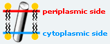
| - | *[[Alpha-bungarotoxin]] is a nicotinic cholinergic antagonist that is found within the venom of ''Bungarus multicinctus'', a South-asian snake belonging to a group commonly known as kraits. | + | The receptor is a transmembrane pentameric glycoprotein. It cylindrical in appearance by electron microscopy approximately 16nm in length and 8nm in diameter. The main ion channel is composed of a water pore that runs through the entire length of the protein. If viewed from the synaptic cleft, the protein will look like a pseudo-symmetrical rosette shown in the picture below composed of 10 different alpha and 4 different beta subunits. |
| | + | *<scene name='58/584302/Cv/1'>Side view</scene>. |
| | + | *<scene name='58/584302/Cv/2'>View from extracellular side</scene>. |
| | + | *<scene name='58/584302/Cv/3'>View from cytoplasmic side</scene>. |
| | + | |
| | + | *[[Alpha-bungarotoxin]] is a nicotinic cholinergic antagonist that is found within the venom of ''Bungarus multicinctus'', a South-asian snake. |
| | *[[Binding site of AChR]] | | *[[Binding site of AChR]] |
| | *[[Acetylcholine Receptor and its Reaction to Cobra Venom]] | | *[[Acetylcholine Receptor and its Reaction to Cobra Venom]] |
| | + | When cobra venom is introduced into the body is moves along the bloodstream to a diaphragm muscle. It works as a postsynaptic neurotoxin binding to the receptor as an extracellular ligand by interacting with OH group leaving the acetylcholine channel open which releases ions used in creating an action potential. There must be 5 molecules of cobra toxin (red) to block the receptor (blue) as each molecule binds with an individual alpha chain on the acetylcholine receptor. The 2nd image depicts an individual toxin binding with one chain on the receptor, both in the same color. <scene name='77/778333/Cobra_snake_venom/3'>Cobra Venom Interaction with Acetylcholine Receptor</scene>. This representation shows each molecule of the <scene name='77/778333/Venom_receptor_piece/1'>Cobra toxin binding to one chain of the receptor</scene>. |
| | | | |
| | ==Anionic cys-loop receptors== | | ==Anionic cys-loop receptors== |
| | ===GABA<sub>A</sub> receptors=== | | ===GABA<sub>A</sub> receptors=== |
| - | *[[4cof]] – hGABAA subunit β-3 - human<br />
| + | |
| - | *[[6i53]], [[6hup]], [[6huo]], [[6huk]], [[6huj]], [[6hug]] – hGABAA subunits β-3 + α-1+γ-2 + megabody – Cryo EM<br />
| + | See [[GABAA receptor]] |
| - | *[[6d6u]], [[6d6t]] – hGABAA subunits β-3 +α-1+γ-2 + antibody + GABA + flumazenil – Cryo EM<br />
| + | |
| - | *[[6a96]] – hGABAA subunits β-3 +α-5 + nanobody + GABA – Cryo EM<br />
| + | |
| - | *[[6hsn]] – rGABAA subunit α-3 + gephyrin + ADP - rat<br />
| + | |
| - | *[[6dw1]], [[6dw0]] – rGABAA subunits β-1 +α-1+γ-2 + GABA – Cryo EM<br />
| + | |
| | | | |
| | =[[Ionotropic Glutamate Receptors]]= | | =[[Ionotropic Glutamate Receptors]]= |
| | ==AMPA glutamate receptor== | | ==AMPA glutamate receptor== |
| | *[[Molecular Playground/Glutamate Receptor|AMPA glutamate receptor]] by [http://www.umass.edu/cbi/ University of Massachusetts Amherst Chemistry-Biology Interface Program] at UMass Amherst and on display at the [http://www.molecularplayground.org/ Molecular Playground]. | | *[[Molecular Playground/Glutamate Receptor|AMPA glutamate receptor]] by [http://www.umass.edu/cbi/ University of Massachusetts Amherst Chemistry-Biology Interface Program] at UMass Amherst and on display at the [http://www.molecularplayground.org/ Molecular Playground]. |
| | + | Full view of the glutamate receptor shows the overall structure (N-terminal, ligand-binding and transmembrane domains) in <scene name='User:Mariel_Feliciano/sandbox_1/Full_view_black_background/6'>ribbon</scene> and <scene name='User:Mariel_Feliciano/sandbox_1/Full_view_spacefill/2'>spacefilling</scene> models. <scene name='User:Mariel_Feliciano/sandbox_1/Amino_terminal_domains/2'>N-terminal domain</scene> is a part of the extracellular domain. This domain is implicated in receptor assembly, trafficking, and localization. |
| | + | *<scene name='Molecular_Playground/Glutamate_Receptor/Transmembrane_domains/5'>Transmembrane Domain</scene>. |
| | + | *<scene name='Molecular_Playground/Glutamate_Receptor/Transmembrane_domains_pore2/1'>Transmembrane Domain, other representaion</scene>. This domain widens in response to glutamate binding allowing for positive ions to pass through the post-synaptic membrane. |
| | + | *<scene name='Molecular_Playground/Glutamate_Receptor/Glu_antagoinist/2'>Receptor antagonist 2K200225 binding site</scene>. |
| | + | *<scene name='Molecular_Playground/Glutamate_Receptor/Glu_agonist_/2'>Glutamate binding site</scene>. |
| | *[[Glutamate receptor (GluA2)]] | | *[[Glutamate receptor (GluA2)]] |
| | The homomeric rat GluA2 receptor <scene name='User:Wayne_Decatur/Sandbox_Glutamate_receptor/Default3kg2/1'>has 4 subunits</scene> arranged in a 'Y'-shape with the <scene name='User:Wayne_Decatur/Sandbox_Glutamate_receptor/Meas3kg2/1'> 'top' being about 3 times the width of the 'bottom'</scene>. This structure is a functional homotetramer of the AMPA-subtype; native ionotropic glutamate receptors are almost exclusively heterotetramers. {{Link Toggle FancyCartoonHighQualityView}}. | | The homomeric rat GluA2 receptor <scene name='User:Wayne_Decatur/Sandbox_Glutamate_receptor/Default3kg2/1'>has 4 subunits</scene> arranged in a 'Y'-shape with the <scene name='User:Wayne_Decatur/Sandbox_Glutamate_receptor/Meas3kg2/1'> 'top' being about 3 times the width of the 'bottom'</scene>. This structure is a functional homotetramer of the AMPA-subtype; native ionotropic glutamate receptors are almost exclusively heterotetramers. {{Link Toggle FancyCartoonHighQualityView}}. |
| Line 112: |
Line 119: |
| | '''Inward rectifier KCh:''' | | '''Inward rectifier KCh:''' |
| | | | |
| - | **[[6c3p]], [[6c3o]] - hIRK 11 + SUR1 + ATP + ADP – Cryo EM<br />
| + | See [[Potassium channel 3D structures]] |
| - | **[[6baa]] - hIRK 11 + SUR1 + ATP + diabetes drug – Cryo EM<br />
| + | |
| - | **[[3ukm]] – hIRK TWIK-1<br />
| + | |
| - | **[[3um7]] – hIRK TRAAK<br />
| + | |
| - | **[[4ruf]], [[4rue]] - hIRK TRAAK (mutant)<br />
| + | |
| - | **[[4wfh]], [[4wfg]], [[4wfe]], [[4wff]] - hIRK TRAAK + antibody<br />
| + | |
| - | **[[4i9w]] - hIRK TRAAK (mutant) + antibody<br />
| + | |
| - | **[[6pz9]], [[6pza]], [[5twv]] - rIRK 11 + SUR1 + ATP + diabetes drug – Cryo EM<br />
| + | |
| - | **[[1u4f]],[[3agw]] - mIRK 2 cytoplasmic domain<br />
| + | |
| - | **[[2xky]] - mIRK 2 cytoplasmic domain - EM<br />
| + | |
| - | **[[2gix]], [[3vsq]] - mIRK 2 cytoplasmic domain (mutant)<br />
| + | |
| - | **[[2e4f]] - mIRK 2 fragment<br />
| + | |
| - | **[[1n9p]], [[1u4e]] - mIRK 1 cytoplasmic domain<br />
| + | |
| - | **[[3k6n]] - mIRK 1 cytoplasmic domain (mutant)<br />
| + | |
| - | **[[5um4]] - mIRK 3.1 (mutant)<br />
| + | |
| - | **[[3at8]], [[3at9]], [[3ata]], [[3atb]], [[3atd]], [[3ate]], [[3atf]], [[3auw]], [[3syo]] - mIRK 3.2<br />
| + | |
| - | **[[6xis]], [[6xit]] - mIRK 3.2 – Cryo EM<br />
| + | |
| - | **[[3syc]], [[3syp]] - mIRK 3.2 (mutant) <br />
| + | |
| - | **[[3sya]] - mIRK 3.2 + PIP2<br />
| + | |
| - | **[[3syq]] - mIRK 3.2 (mutant) + PIP2<br />
| + | |
| - | **[[4kfm]] - mIRK 3.2 + guanine nucleotide-biding protein<br />
| + | |
| - | **[[5wua]], [[5twv]] - mIRK 11 + SUR1 - Cryo EM<br />
| + | |
| - | **[[5ywc]], [[5ywb]] - mIRK 11 + SUR1 + ADP – Cryo EM<br />
| + | |
| - | **[[5ywa]], [[5yw9]], [[5yw8]] - mIRK 11 + SUR1 + ATP – Cryo EM<br />
| + | |
| - | **[[5ykg]], [[5ykf]], [[5yke]], [[6jb1]] - mIRK 11 + SUR1 + ATP + diabetes drug – Cryo EM<br />
| + | |
| - | **[[6m85]] – cIRK 2.2<br />
| + | |
| - | **[[3spj]], [[6m86]] – cIRK 2.2 (mutant)<br />
| + | |
| - | **[[3spc]] – cIRK 2.2 + DGPP <br />
| + | |
| - | **[[3spg]], [[3sph]], [[5kum]], [[5kuk]], [[6m84]] – cIRK 2.2 (mutant) + PIP2<br />
| + | |
| - | **[[3spi]] – cIRK 2.2 + PIP2<br />
| + | |
| - | **[[7cal]] - AtIRK KAT1 – Cryo EM<br />
| + | |
| - | **[[1xl4]], [[1xl6]], [[2wlk]], [[2x6a]], [[2x6b]], [[2x6c]], [[6o9u]] - MmIRK KIRBAC3.1<br />
| + | |
| - | **[[4lp8]], [[6o9t]], [[6o9v]] - MmIRK KIRBAC3.1 (mutant) <br />
| + | |
| - | **[[3zrs]] – MmIRK 10<br />
| + | |
| - | **[[1p7b]] - BpIRK C-terminal<br />
| + | |
| - | | + | |
| | | | |
| | </StructureSection> | | </StructureSection> |
| | ==References== | | ==References== |
| | <references/> | | <references/> |
| | + | [[Category:Topic Page]] |
| See also Receptor.
Cys-loop receptors
Cationic cys-loop receptors
Serotonin type-3 receptor (5-HT3-R)
is a pentameric cation-selective ion channel and plays a role in neuronal excitation to release neurotransmitters from the postsynaptic neuron. Opening of the cation channel causes an influx of sodium and calcium through the receptor pore leading to a membrane depolarization. Five receptor subunits, A to E, have been found in humans but only subunits A and B have been found in rodents. When experimentally expressed in a host, the 5-HT3 receptor is comprised of either A or AB subunits which can result in a homopentameric receptor or a heteropentameric receptor respectively. The A and B subunits are found throughout the brain in areas such as the hippocampus and amygdala. 5-HT3 is a transmembrane channel that is stimulated to open state by the interaction of the receptor with serotonin in the extracellular space. The binding site is comprised of 6 loops from 2 adjacent subunits in the extracellular N-terminal domain. Loops A, B and C form the principal subunit and contain the N128, W183 and Y234. Loops D, E and F form the complementary subunit of the binding site and contain the important side chains W90, Y143 and W195. The transmembrane region is comprised of multiple alpha helical structures and mediates ion flow and ion specificity.
The serotonin type-3 receptor is a cation selective transmembrane protein channel that belongs to the Cys–loop Ligand-Gated Ion Channel (LGIC) superfamily, which also includes receptors for nicotinic acetylcholine (, PDB code 2bg9), γ-aminobutyric acid and glycine. 5-HT3-R is involved in signal transmission in the central and peripheral nervous system and its malfunctioning leads to neurodegenerative and psychiatric diseases, therefore it is an important target for drug design research. A few drugs active against 5-HT3-R are already on the market, such as, for example, palonosetron (http://en.wikipedia.org/wiki/Palonosetron) and granisetron (http://en.wikipedia.org/wiki/Granisetron).
The 5-HT3R is made of 5 monomers assembled in a to form an ion channel permeable to small ions (Na+, K+); each subunit contains 3 domains: an (shown on the example of nAChR, 2bg9). To date, 5 different 5-HT3-R subunits have been identified, the 5-HT3 A, B, C, D and E; however, only subunits A and B have been extensively characterized experimentally. The of nAChR is located at the extracellular region, at the interface between 2 monomers (α-γ and α-δ; 2 identical α monomers, chains A and D, are colored in same color - lavender), called the principal and the complementary subunits.
The 3D structure of 5-HT3-R has not been experimentally solved yet; however, it has been obtained computationally by means of homology modelling techniques. (http://salilab.org/modeller/)
Thus, the are modelled by homology with the 3D structure of the nAChR subunit A (2bg9-A) and are used to assemble receptor structures as pseudo-symmetric pentamers made either of or of in a still debated arrangement.[2] Subunits A and B are colored in magenta and red, respectively.
A complete characterization of the extracellular moiety of the (AA dimer is shown, principal subunit is colored in cyan and complementary is in blue, is obtained by the Computational Alanine Scanning Mutagenesis (CASM) approach [3], which simulates the substitution, one by one, of all the amino acid residues at the subunit-subunit interfaces with an Ala, thus to assess the interface binding contribution of single residue side-chains. The are classified as “hot spots” that stabilize the interface by more than 4 kcal/mol and “warm spots” that contribute to interface stabilization by more than 2 kcal/mol. Interface residues are shown in spacefill representation, hot spot residues are colored in red and warm spots residues are are in orange.
From this analysis the located at the interface core and formed by residues W178 (principal subunit), Y68, Y83, W85 and Y148 (complementary subunit) is highlighted.[4] In addition, 2 important groups of interface residues are probably involved in the coupling of and binding to channel activation/inactivation: W116-H180-L179-W178-E124-F125 (principal subunit) and Y136-Y138-Y148-W85-(P150) (complementary subunit), where W178 and Y148 appear to be critical residues for the binding/activation mechanism. Finally, the (principal subunit of AA is colored in cyan, principal subunit BB is colored in darkmagenta, complementary subunit AA is in blue and complementary subunit BB is in magenta) shows differences which could explain the reasons why the homopentamer 5-HT3B-R, if expressed, is not functional.[5]
The receptor is bullet-shaped and consists of 5 subunits (A-E) that form an oligomer. In the center of this pentamer of is a ligand-gated ion channel full of water, which the 5 subunits enclose pseudo-symmetrically. Each subunit of the 5-HT3 receptor consists of 3 regions; the extracellular region, the transmembrane region, and the intracellular region. The is relatively large compared to the other 2 regions, and contains a short C-terminus and a larger N-terminus. The N-terminus of the extracellular region is where the ligand binding occurs, and therefore deals with the agonists and antagonists. These are located between 2 bordering subunits, assembled from 3 α-helices of 1 subunit and 3 β-strands from the other subunit. Such connection creates a binding pocket with a small number of residues from each subunit pointed into the binding pocket, as opposed to the large number of residues that are pointing from the binding pocket. This binding pocket shrinks around agonists, encapsulating them, and widens around antagonists, repulsing them. The is within the C-terminus region, and contains 4 α-helical domains (M1-M4) that stretch the length of this inner, transmembrane area. These 4 α-helical domains conduct the channel openings via ion selectivity, depending on both charge and size. M2, the porous domain, contains rings of charged amino acids at both its start and its , accounting for M2 main contribution to ion selectivity. The M3 and M4 α-helices create a large with one another, thus assembling the .
Nicotinic acetylcholine receptors
The receptor is a transmembrane pentameric glycoprotein. It cylindrical in appearance by electron microscopy approximately 16nm in length and 8nm in diameter. The main ion channel is composed of a water pore that runs through the entire length of the protein. If viewed from the synaptic cleft, the protein will look like a pseudo-symmetrical rosette shown in the picture below composed of 10 different alpha and 4 different beta subunits.
When cobra venom is introduced into the body is moves along the bloodstream to a diaphragm muscle. It works as a postsynaptic neurotoxin binding to the receptor as an extracellular ligand by interacting with OH group leaving the acetylcholine channel open which releases ions used in creating an action potential. There must be 5 molecules of cobra toxin (red) to block the receptor (blue) as each molecule binds with an individual alpha chain on the acetylcholine receptor. The 2nd image depicts an individual toxin binding with one chain on the receptor, both in the same color. . This representation shows each molecule of the .
Anionic cys-loop receptors
GABAA receptors
See GABAA receptor
AMPA glutamate receptor
Full view of the glutamate receptor shows the overall structure (N-terminal, ligand-binding and transmembrane domains) in and models. is a part of the extracellular domain. This domain is implicated in receptor assembly, trafficking, and localization.
- .
- . This domain widens in response to glutamate binding allowing for positive ions to pass through the post-synaptic membrane.
- .
- .
- Glutamate receptor (GluA2)
The homomeric rat GluA2 receptor arranged in a 'Y'-shape with the . This structure is a functional homotetramer of the AMPA-subtype; native ionotropic glutamate receptors are almost exclusively heterotetramers. .
Domains
The subunits themselves are modular [6]and the major domains are found in layers in the tetrameric structure.
- The 'top' layer is composed of the
- This .
- participates directly in agonist/competitive antagonist binding, affects activation gating, and is the portion that forms the 'middle' layer.
- in the structure.
- The [7], was studied as a treatment for stroke because it had demonstrated neuroprotective efficacy in experimental models of stroke and tolerability in healthy volunteers; however, in a multicenter, double-blind, randomized, placebo-controlled phase II trial, it was found to have significant sedative effects in patients with acute stroke which precludes its further development as a neuroprotective agent[8].
- is the portion that forms the membrane-spanning on the 'bottom' of the solved structure.
- To help give a better idea of how the glutamate receptor is oriented on the cell surface in the membrane lipid bilayer, as calculated by the Orientations of Proteins in Membranes database (University of Michigan, USA) is shown with the red patch of spheres indicating the boundary of the hydrophobic core closest to the outside of the cell and the dark blue patch of spheres indicating the boundary closest to the inside of the cell.
- The carboxy-terminal domain that plays a role in both receptor localization and regulation is not seen in the structure but would be below the transmembrane domain as it is cytoplasmic.
Domain swapping between the subunits and symmetry mismatch between the domains
- Unanticipated is the domain swapping and crossover that occurs between the subunits interactions. In order to discuss the remarkable swapping, it is best to :
A B C D
- Considering each chain, there is crossover as the pairs of subunits seen in the ATD are swapped in the LBD.
- In the ATD domain -
- .
- And the .
- While that is going on, in the ATD there is also inter-pair interactions mediated between . Note this view really highlights the two-fold symmetry between the A-B and C-D pairs at the level of the ATD.
- In the LBD domain -
- Whereas in the ATD domain A and B paired up, in the LBD.
- And the .
- While that is going on, in the LBD there is also extensive inter-pair interactions mediated between . Note this view highlights the two-fold symmetry between the A-D and B-C pairs at the level of the LBD. .
- The domain swapping can be observed from the side following the backbone of each chain as well: , , , and . And .
- The . Thus, remarkably, the symmetry switches from an overall two-fold symmetry for the ATD and LBD to four-fold for the TMD.
As a result of the swapping and symmetry mismatch, there is subunit non-equivalence; even though all the chains are the same chemically, there are 2 distinct conformations of the subunits. This means there are 2 matching pairs of subunits.
-
-
- Subunit A is equivalent to Subunit C (in the small structure window in this section). In the main window, a .
- Subunit B is equivalent to Subunit D (in the small structure window in this section). In the main window, a .
However, each of the subunit A/C group though is distinct from those of the B/D group. Having established the two equivalent groups we can simplify the discussion of the relationship between the two pairs by focusing solely on comparing Subunit A' and Subunit B.
The domains themselves stay relatively static between the two conformational forms, with the linkers in between and the resulting arrangement changing. This is best illustrated by superposition of the individual domains of Subunit A and Subunit B:
between the two conformational forms.
- The linkers are key; besides playing roles in domain swapping and resolving the symmetry mismatch, they are also responsible for relaying the modulation signals from the ATD to the other domains and signaling the conformational change of the LBD to control the opening and closing of the pore. Beyond the two conformations seen here though this particular structure (3kg2) of the receptor does not shed light on the transduction process.
Transmembrane domain architecture and the occluded pore
-
- The segments shown again, .
- There is consistent with the channel being in a closed state with the antagonist ZK200775 bound to the LBD.
- It is that occludes the channel. [BE PATIENT as a small surface is generated.]
- Note . This is in part is why the symmetry is only approximately four-fold and is one of the several intriguing observations in regard to symmetry for this macromolecule. In fact, the location of 2-fold symmetry at the ends of M3 is just above the portion that spans the membrane and is close to the last region of the structure that doesn't show four-fold symmetry as abruptly below this point everything is 4-fold symmetric.
- To better observe the contributions of each of the membrane segments to the subunit-subunit interactions, . [Note: this scene generates a substantial surface which may take about a minute to calculate. Be patient.]
- Note that the M4 segment associates with the ion-channel core of an adjacent subunit.
- .
- The TMD domain of the GluA2 receptor shares structural and sequence similarity with the pore region of the potassium (K+), as hinted at by earlier work[9][10][11]. Here the pore region of Streptomyces lividans potassium channel (1bl8), specifically the . The of the K+ channel even though these portions weren't even included in the calculation of the alignment seen here.
ATP-gated P2X receptor cation channel family
Examples:
3h9v, 3i5d
PIP2-gated channels
Phosphatidylinositol 4,5-bisphosphate (PIP2) binds to and directly activates inwardly rectifying potassium channels.
Inward rectifier KCh:
See Potassium channel 3D structures
|

