1fxx
From Proteopedia
(Difference between revisions)
| Line 3: | Line 3: | ||
<StructureSection load='1fxx' size='340' side='right'caption='[[1fxx]], [[Resolution|resolution]] 2.40Å' scene=''> | <StructureSection load='1fxx' size='340' side='right'caption='[[1fxx]], [[Resolution|resolution]] 2.40Å' scene=''> | ||
== Structural highlights == | == Structural highlights == | ||
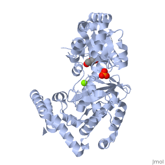
| - | <table><tr><td colspan='2'>[[1fxx]] is a 1 chain structure with sequence from [https://en.wikipedia.org/wiki/ | + | <table><tr><td colspan='2'>[[1fxx]] is a 1 chain structure with sequence from [https://en.wikipedia.org/wiki/Escherichia_coli Escherichia coli]. Full crystallographic information is available from [http://oca.weizmann.ac.il/oca-bin/ocashort?id=1FXX OCA]. For a <b>guided tour on the structure components</b> use [https://proteopedia.org/fgij/fg.htm?mol=1FXX FirstGlance]. <br> |
| - | </td></tr><tr id='ligand'><td class="sblockLbl"><b>[[Ligand|Ligands:]]</b></td><td class="sblockDat" id="ligandDat"><scene name='pdbligand=GOL:GLYCEROL'>GOL</scene>, <scene name='pdbligand=MG:MAGNESIUM+ION'>MG</scene>, <scene name='pdbligand=PO4:PHOSPHATE+ION'>PO4</scene | + | </td></tr><tr id='method'><td class="sblockLbl"><b>[[Empirical_models|Method:]]</b></td><td class="sblockDat" id="methodDat">X-ray diffraction, [[Resolution|Resolution]] 2.4Å</td></tr> |
| - | + | <tr id='ligand'><td class="sblockLbl"><b>[[Ligand|Ligands:]]</b></td><td class="sblockDat" id="ligandDat"><scene name='pdbligand=GOL:GLYCEROL'>GOL</scene>, <scene name='pdbligand=MG:MAGNESIUM+ION'>MG</scene>, <scene name='pdbligand=PO4:PHOSPHATE+ION'>PO4</scene></td></tr> | |
<tr id='resources'><td class="sblockLbl"><b>Resources:</b></td><td class="sblockDat"><span class='plainlinks'>[https://proteopedia.org/fgij/fg.htm?mol=1fxx FirstGlance], [http://oca.weizmann.ac.il/oca-bin/ocaids?id=1fxx OCA], [https://pdbe.org/1fxx PDBe], [https://www.rcsb.org/pdb/explore.do?structureId=1fxx RCSB], [https://www.ebi.ac.uk/pdbsum/1fxx PDBsum], [https://prosat.h-its.org/prosat/prosatexe?pdbcode=1fxx ProSAT]</span></td></tr> | <tr id='resources'><td class="sblockLbl"><b>Resources:</b></td><td class="sblockDat"><span class='plainlinks'>[https://proteopedia.org/fgij/fg.htm?mol=1fxx FirstGlance], [http://oca.weizmann.ac.il/oca-bin/ocaids?id=1fxx OCA], [https://pdbe.org/1fxx PDBe], [https://www.rcsb.org/pdb/explore.do?structureId=1fxx RCSB], [https://www.ebi.ac.uk/pdbsum/1fxx PDBsum], [https://prosat.h-its.org/prosat/prosatexe?pdbcode=1fxx ProSAT]</span></td></tr> | ||
</table> | </table> | ||
== Function == | == Function == | ||
| - | + | [https://www.uniprot.org/uniprot/EX1_ECOLI EX1_ECOLI] Also functions as a DNA deoxyribophosphodiesterase that releases deoxyribose-phosphate moieties following the cleavage DNA at an apurinic/apyrimidinic (AP) site by either an AP endonuclease AP lyase. | |
== Evolutionary Conservation == | == Evolutionary Conservation == | ||
[[Image:Consurf_key_small.gif|200px|right]] | [[Image:Consurf_key_small.gif|200px|right]] | ||
| Line 20: | Line 20: | ||
</jmol>, as determined by [http://consurfdb.tau.ac.il/ ConSurfDB]. You may read the [[Conservation%2C_Evolutionary|explanation]] of the method and the full data available from [http://bental.tau.ac.il/new_ConSurfDB/main_output.php?pdb_ID=1fxx ConSurf]. | </jmol>, as determined by [http://consurfdb.tau.ac.il/ ConSurfDB]. You may read the [[Conservation%2C_Evolutionary|explanation]] of the method and the full data available from [http://bental.tau.ac.il/new_ConSurfDB/main_output.php?pdb_ID=1fxx ConSurf]. | ||
<div style="clear:both"></div> | <div style="clear:both"></div> | ||
| - | <div style="background-color:#fffaf0;"> | ||
| - | == Publication Abstract from PubMed == | ||
| - | Exonuclease I (ExoI) from Escherichia coli is a monomeric enzyme that processively degrades single stranded DNA in the 3' to 5' direction and has been implicated in DNA recombination and repair. Determination of the structure of ExoI to 2.4 A resolution using X-ray crystallography verifies the expected correspondence between a region of ExoI and the exonuclease (or proofreading) domains of the DNA polymerases. The overall fold of ExoI also includes two other regions, one of which extends the exonuclease domain and another that can be described as an elaborated SH3 domain. These three regions combine to form a molecule that is shaped like the letter C, although it also contains a segment that effectively converts the C into an O-like shape. The structure of ExoI thus provides additional support for the idea that DNA metabolizing enzymes achieve processivity by completely enclosing the DNA. | ||
| - | |||
| - | Structure of Escherichia coli exonuclease I suggests how processivity is achieved.,Breyer WA, Matthews BW Nat Struct Biol. 2000 Dec;7(12):1125-8. PMID:11101894<ref>PMID:11101894</ref> | ||
| - | |||
| - | From MEDLINE®/PubMed®, a database of the U.S. National Library of Medicine.<br> | ||
| - | </div> | ||
| - | <div class="pdbe-citations 1fxx" style="background-color:#fffaf0;"></div> | ||
==See Also== | ==See Also== | ||
*[[Exonuclease|Exonuclease]] | *[[Exonuclease|Exonuclease]] | ||
*[[Exonuclease 3D structures|Exonuclease 3D structures]] | *[[Exonuclease 3D structures|Exonuclease 3D structures]] | ||
| - | == References == | ||
| - | <references/> | ||
__TOC__ | __TOC__ | ||
</StructureSection> | </StructureSection> | ||
| - | [[Category: | + | [[Category: Escherichia coli]] |
| - | + | ||
[[Category: Large Structures]] | [[Category: Large Structures]] | ||
| - | [[Category: Breyer | + | [[Category: Breyer WA]] |
| - | [[Category: Matthews | + | [[Category: Matthews BW]] |
| - | + | ||
| - | + | ||
| - | + | ||
| - | + | ||
Current revision
THE STRUCTURE OF EXONUCLEASE I SUGGESTS HOW PROCESSIVITY IS ACHIEVED
| |||||||||||