We apologize for Proteopedia being slow to respond. For the past two years, a new implementation of Proteopedia has been being built. Soon, it will replace this 18-year old system. All existing content will be moved to the new system at a date that will be announced here.
FirstGlance/How To Measure A Virus Capsid
From Proteopedia
(Difference between revisions)
| (7 intermediate revisions not shown.) | |||
| Line 1: | Line 1: | ||
<StructureSection load='' size='350' side='right' caption='' scene=''> | <StructureSection load='' size='350' side='right' caption='' scene=''> | ||
| - | <table style="background-color:#ffffa0;" class='wikitable'><tr><td> | ||
| - | |||
| - | This page describes the use of FirstGlance in Jmol version 4.0. Its release is expected soon, but it is not yet publicly available. [[User:Eric Martz|Eric Martz]] 22:25, 26 July 2022 (UTC) | ||
| - | |||
| - | </td></tr></table> | ||
| - | |||
{{Template:EEEV}} | {{Template:EEEV}} | ||
| - | Quick Start: [http:// | + | '''Quick Start''': [http://firstglance.jmol.org/fg.htm?mol=6mx4 Analyze the EEEV capsid 6mx4 in FirstGlance]. |
[[FirstGlance in Jmol]] automatically constructs the capsid, and simplifies it to a subset of alpha carbon atoms small enough (not more than 25,000) to be analyzed efficiently in FirstGlance and [[JSmol]], both of which run in the Javascript of the web browser. FirstGlance offers a number of useful color schemes, including distance from center, colors that distinguish sequence-identical groups of chains, and [[Help:Color_Keys#Rainbows:_N_to_C.2C_5.27_to_3.27|amino-to-carboxy rainbow]]. When the structure is small enough that all alpha carbons can be displayed (not more than 250,000 alpha carbons; EEEV has 242,340), it can also be colored by charge, hydrophobic vs. polar, or [[evolutionary conservation]]. | [[FirstGlance in Jmol]] automatically constructs the capsid, and simplifies it to a subset of alpha carbon atoms small enough (not more than 25,000) to be analyzed efficiently in FirstGlance and [[JSmol]], both of which run in the Javascript of the web browser. FirstGlance offers a number of useful color schemes, including distance from center, colors that distinguish sequence-identical groups of chains, and [[Help:Color_Keys#Rainbows:_N_to_C.2C_5.27_to_3.27|amino-to-carboxy rainbow]]. When the structure is small enough that all alpha carbons can be displayed (not more than 250,000 alpha carbons; EEEV has 242,340), it can also be colored by charge, hydrophobic vs. polar, or [[evolutionary conservation]]. | ||
| Line 14: | Line 8: | ||
==Slab of the Capsid== | ==Slab of the Capsid== | ||
In order to estimate the dimensions of the capsid, it is useful to "cut out a slab". | In order to estimate the dimensions of the capsid, it is useful to "cut out a slab". | ||
| - | + | {{Template:EEEV-slab}} | |
| - | + | ||
| - | + | ||
| - | + | ||
| - | + | ||
| - | + | ||
| - | + | ||
| - | + | ||
| - | + | ||
| - | + | ||
| - | + | ||
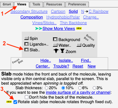
To obtain this slab in FirstGlance, you simply depress a ''Slab'' button. By default, the thickness of the slab is 10% of the diameter of the capsid (but this is adjustable). Here is a '''snapshot of the ''Views'' tab of FirstGlance'''. After the initial view of the capsid appears, getting this slab takes 3 clicks indicated by the red arrows: Click on the ''Views'' tab, depress the ''Slab'' button, and check ''Rotate slab''. | To obtain this slab in FirstGlance, you simply depress a ''Slab'' button. By default, the thickness of the slab is 10% of the diameter of the capsid (but this is adjustable). Here is a '''snapshot of the ''Views'' tab of FirstGlance'''. After the initial view of the capsid appears, getting this slab takes 3 clicks indicated by the red arrows: Click on the ''Views'' tab, depress the ''Slab'' button, and check ''Rotate slab''. | ||
| Line 55: | Line 39: | ||
</jmol> | </jmol> | ||
(total 720 protein chains). | (total 720 protein chains). | ||
| + | |||
| + | For more about the EEEV capsid, see [[FirstGlance/Virus_Capsids_and_Other_Large_Assemblies#Eastern_Equine_Encephalitis_Virus|Virus Capsids and Other Large Assemblies]]. | ||
</StructureSection> | </StructureSection> | ||
| + | |||
| + | ==Reference== | ||
| + | <references /> | ||
Current revision
| |||||||||||
Reference
- ↑ Hasan SS, Sun C, Kim AS, Watanabe Y, Chen CL, Klose T, Buda G, Crispin M, Diamond MS, Klimstra WB, Rossmann MG. Cryo-EM Structures of Eastern Equine Encephalitis Virus Reveal Mechanisms of Virus Disassembly and Antibody Neutralization. Cell Rep. 2018 Dec 11;25(11):3136-3147.e5. doi: 10.1016/j.celrep.2018.11.067. PMID:30540945 doi:http://dx.doi.org/10.1016/j.celrep.2018.11.067

