We apologize for Proteopedia being slow to respond. For the past two years, a new implementation of Proteopedia has been being built. Soon, it will replace this 18-year old system. All existing content will be moved to the new system at a date that will be announced here.
1a2e
From Proteopedia
(Difference between revisions)
| Line 4: | Line 4: | ||
== Structural highlights == | == Structural highlights == | ||
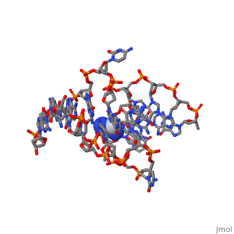
<table><tr><td colspan='2'>[[1a2e]] is a 2 chain structure. Full crystallographic information is available from [http://oca.weizmann.ac.il/oca-bin/ocashort?id=1A2E OCA]. For a <b>guided tour on the structure components</b> use [https://proteopedia.org/fgij/fg.htm?mol=1A2E FirstGlance]. <br> | <table><tr><td colspan='2'>[[1a2e]] is a 2 chain structure. Full crystallographic information is available from [http://oca.weizmann.ac.il/oca-bin/ocashort?id=1A2E OCA]. For a <b>guided tour on the structure components</b> use [https://proteopedia.org/fgij/fg.htm?mol=1A2E FirstGlance]. <br> | ||
| - | </td></tr><tr id='ligand'><td class="sblockLbl"><b>[[Ligand|Ligands:]]</b></td><td class="sblockDat" id="ligandDat"><scene name='pdbligand=CPT:CISPLATIN'>CPT</scene></td></tr> | + | </td></tr><tr id='method'><td class="sblockLbl"><b>[[Empirical_models|Method:]]</b></td><td class="sblockDat" id="methodDat">X-ray diffraction, [[Resolution|Resolution]] 1.63Å</td></tr> |
| + | <tr id='ligand'><td class="sblockLbl"><b>[[Ligand|Ligands:]]</b></td><td class="sblockDat" id="ligandDat"><scene name='pdbligand=CPT:CISPLATIN'>CPT</scene></td></tr> | ||
<tr id='resources'><td class="sblockLbl"><b>Resources:</b></td><td class="sblockDat"><span class='plainlinks'>[https://proteopedia.org/fgij/fg.htm?mol=1a2e FirstGlance], [http://oca.weizmann.ac.il/oca-bin/ocaids?id=1a2e OCA], [https://pdbe.org/1a2e PDBe], [https://www.rcsb.org/pdb/explore.do?structureId=1a2e RCSB], [https://www.ebi.ac.uk/pdbsum/1a2e PDBsum], [https://prosat.h-its.org/prosat/prosatexe?pdbcode=1a2e ProSAT]</span></td></tr> | <tr id='resources'><td class="sblockLbl"><b>Resources:</b></td><td class="sblockDat"><span class='plainlinks'>[https://proteopedia.org/fgij/fg.htm?mol=1a2e FirstGlance], [http://oca.weizmann.ac.il/oca-bin/ocaids?id=1a2e OCA], [https://pdbe.org/1a2e PDBe], [https://www.rcsb.org/pdb/explore.do?structureId=1a2e RCSB], [https://www.ebi.ac.uk/pdbsum/1a2e PDBsum], [https://prosat.h-its.org/prosat/prosatexe?pdbcode=1a2e ProSAT]</span></td></tr> | ||
</table> | </table> | ||
| - | <div style="background-color:#fffaf0;"> | ||
| - | == Publication Abstract from PubMed == | ||
| - | cis-diamminedichloroplatinum (II) (cisplatin) is a powerful anti-tumor drug whose target is cellular DNA. In the reaction between DNA and cisplatin, covalent intrastrand and interstrand cross-links (ICL) are formed. Two solution structures of the ICL have been published recently. In both models the double-helix is bent and unwound but with significantly different angle values. We solved the crystal structure at 100K of a double-stranded DNA decamer containing a single cisplatin ICL, using the anomalous scattering (MAD) of platinum as a unique source of phase information. We found 47 degrees for double-helix bending and 70 degrees for unwinding in agreement with previous electrophoretic assays. The crystals are stabilized by intermolecular contacts involving two cytosines extruded from the double-helix, one of which makes a triplet with a terminal G.C pair. The platinum coordination is nearly square and the platinum residue is embedded into a cage of nine water molecules linked to the cross-linked guanines, to the two amine groups, and to the phosphodiester backbone through other water molecules. This water molecule organization is discussed in relation with the chemical stability of the ICL. | ||
| - | |||
| - | Crystal structure of a double-stranded DNA containing a cisplatin interstrand cross-link at 1.63 A resolution: hydration at the platinated site.,Coste F, Malinge JM, Serre L, Shepard W, Roth M, Leng M, Zelwer C Nucleic Acids Res. 1999 Apr 15;27(8):1837-46. PMID:10101191<ref>PMID:10101191</ref> | ||
| - | |||
| - | From MEDLINE®/PubMed®, a database of the U.S. National Library of Medicine.<br> | ||
| - | </div> | ||
| - | <div class="pdbe-citations 1a2e" style="background-color:#fffaf0;"></div> | ||
| - | == References == | ||
| - | <references/> | ||
__TOC__ | __TOC__ | ||
</StructureSection> | </StructureSection> | ||
Current revision
CRYSTAL STRUCTURE OF A DOUBLE-STRANDED DNA DECAMER CONTAINING A CISPLATIN INTERSTRAND CROSS-LINK ADDUCT
| |||||||||||