We apologize for Proteopedia being slow to respond. For the past two years, a new implementation of Proteopedia has been being built. Soon, it will replace this 18-year old system. All existing content will be moved to the new system at a date that will be announced here.
1ovm
From Proteopedia
(Difference between revisions)
| (11 intermediate revisions not shown.) | |||
| Line 1: | Line 1: | ||
| - | [[Image:1ovm.jpg|left|200px]] | ||
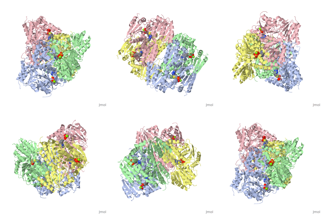
| - | < | + | ==Crystal structure of Indolepyruvate decarboxylase from Enterobacter cloacae== |
| - | + | <StructureSection load='1ovm' size='340' side='right'caption='[[1ovm]], [[Resolution|resolution]] 2.65Å' scene=''> | |
| - | You may | + | == Structural highlights == |
| - | + | <table><tr><td colspan='2'>[[1ovm]] is a 4 chain structure with sequence from [https://en.wikipedia.org/wiki/Enterobacter_cloacae Enterobacter cloacae]. Full crystallographic information is available from [http://oca.weizmann.ac.il/oca-bin/ocashort?id=1OVM OCA]. For a <b>guided tour on the structure components</b> use [https://proteopedia.org/fgij/fg.htm?mol=1OVM FirstGlance]. <br> | |
| - | + | </td></tr><tr id='method'><td class="sblockLbl"><b>[[Empirical_models|Method:]]</b></td><td class="sblockDat" id="methodDat">X-ray diffraction, [[Resolution|Resolution]] 2.65Å</td></tr> | |
| - | + | <tr id='ligand'><td class="sblockLbl"><b>[[Ligand|Ligands:]]</b></td><td class="sblockDat" id="ligandDat"><scene name='pdbligand=MG:MAGNESIUM+ION'>MG</scene>, <scene name='pdbligand=TPP:THIAMINE+DIPHOSPHATE'>TPP</scene></td></tr> | |
| - | + | <tr id='resources'><td class="sblockLbl"><b>Resources:</b></td><td class="sblockDat"><span class='plainlinks'>[https://proteopedia.org/fgij/fg.htm?mol=1ovm FirstGlance], [http://oca.weizmann.ac.il/oca-bin/ocaids?id=1ovm OCA], [https://pdbe.org/1ovm PDBe], [https://www.rcsb.org/pdb/explore.do?structureId=1ovm RCSB], [https://www.ebi.ac.uk/pdbsum/1ovm PDBsum], [https://prosat.h-its.org/prosat/prosatexe?pdbcode=1ovm ProSAT]</span></td></tr> | |
| + | </table> | ||
| + | == Function == | ||
| + | [https://www.uniprot.org/uniprot/DCIP_ENTCL DCIP_ENTCL] | ||
| + | == Evolutionary Conservation == | ||
| + | [[Image:Consurf_key_small.gif|200px|right]] | ||
| + | Check<jmol> | ||
| + | <jmolCheckbox> | ||
| + | <scriptWhenChecked>; select protein; define ~consurf_to_do selected; consurf_initial_scene = true; script "/wiki/ConSurf/ov/1ovm_consurf.spt"</scriptWhenChecked> | ||
| + | <scriptWhenUnchecked>script /wiki/extensions/Proteopedia/spt/initialview01.spt</scriptWhenUnchecked> | ||
| + | <text>to colour the structure by Evolutionary Conservation</text> | ||
| + | </jmolCheckbox> | ||
| + | </jmol>, as determined by [http://consurfdb.tau.ac.il/ ConSurfDB]. You may read the [[Conservation%2C_Evolutionary|explanation]] of the method and the full data available from [http://bental.tau.ac.il/new_ConSurfDB/main_output.php?pdb_ID=1ovm ConSurf]. | ||
| + | <div style="clear:both"></div> | ||
| + | <div style="background-color:#fffaf0;"> | ||
| + | == Publication Abstract from PubMed == | ||
| + | The thiamin diphosphate-dependent enzyme indolepyruvate decarboxylase catalyses the formation of indoleacetaldehyde from indolepyruvate, one step in the indolepyruvate pathway of biosynthesis of the plant hormone indole-3-acetic acid. The crystal structure of this enzyme from Enterobacter cloacae has been determined at 2.65 A resolution and refined to a crystallographic R-factor of 20.5% (Rfree 23.6%). The subunit of indolepyruvate decarboxylase contains three domains of open alpha/beta topology, which are similar in structure to that of pyruvate decarboxylase. The tetramer has pseudo 222 symmetry and can be described as a dimer of dimers. It resembles the tetramer of pyruvate decarboxylase from Zymomonas mobilis, but with a relative difference of 20 degrees in the angle between the two dimers. Active site residues are highly conserved in indolepyruvate/pyruvate decarboxylase, suggesting that the interactions with the cofactor thiamin diphosphate and the catalytic mechanisms are very similar. The substrate binding site in indolepyruvate decarboxylase contains a large hydrophobic pocket which can accommodate the bulky indole moiety of the substrate. In pyruvate decarboxylases this pocket is smaller in size and allows discrimination of larger vs. smaller substrates. In most pyruvate decarboxylases, restriction of cavity size is due to replacement of residues at three positions by large, hydrophobic amino acids such as tyrosine or tryptophan. | ||
| - | + | Crystal structure of thiamindiphosphate-dependent indolepyruvate decarboxylase from Enterobacter cloacae, an enzyme involved in the biosynthesis of the plant hormone indole-3-acetic acid.,Schutz A, Sandalova T, Ricagno S, Hubner G, Konig S, Schneider G Eur J Biochem. 2003 May;270(10):2312-21. PMID:12752451<ref>PMID:12752451</ref> | |
| - | + | ||
| - | + | ||
| - | + | ||
| - | + | ||
| - | + | From MEDLINE®/PubMed®, a database of the U.S. National Library of Medicine.<br> | |
| - | + | </div> | |
| + | <div class="pdbe-citations 1ovm" style="background-color:#fffaf0;"></div> | ||
| - | == | + | ==See Also== |
| - | + | *[[Indole pyruvate decarboxylase|Indole pyruvate decarboxylase]] | |
| + | == References == | ||
| + | <references/> | ||
| + | __TOC__ | ||
| + | </StructureSection> | ||
[[Category: Enterobacter cloacae]] | [[Category: Enterobacter cloacae]] | ||
| - | [[Category: | + | [[Category: Large Structures]] |
| - | + | [[Category: Hubner G]] | |
| - | [[Category: Hubner | + | [[Category: Konig S]] |
| - | [[Category: Konig | + | [[Category: Ricagno S]] |
| - | [[Category: Ricagno | + | [[Category: Sandalova T]] |
| - | [[Category: Sandalova | + | [[Category: Schneider G]] |
| - | [[Category: Schneider | + | [[Category: Schutz A]] |
| - | [[Category: Schutz | + | |
| - | + | ||
| - | + | ||
| - | + | ||
| - | + | ||
| - | + | ||
| - | + | ||
Current revision
Crystal structure of Indolepyruvate decarboxylase from Enterobacter cloacae
| |||||||||||


