We apologize for Proteopedia being slow to respond. For the past two years, a new implementation of Proteopedia has been being built. Soon, it will replace this 18-year old system. All existing content will be moved to the new system at a date that will be announced here.
1pkp
From Proteopedia
(Difference between revisions)
| (11 intermediate revisions not shown.) | |||
| Line 1: | Line 1: | ||
| - | [[Image:1pkp.gif|left|200px]] | ||
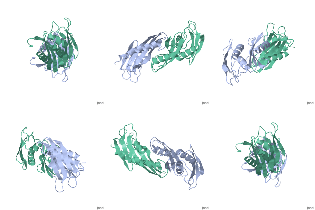
| - | < | + | ==THE STRUCTURE OF RIBOSOMAL PROTEIN S5 REVEALS SITES OF INTERACTION WITH 16S RRNA== |
| - | + | <StructureSection load='1pkp' size='340' side='right'caption='[[1pkp]], [[Resolution|resolution]] 2.80Å' scene=''> | |
| - | + | == Structural highlights == | |
| - | + | <table><tr><td colspan='2'>[[1pkp]] is a 1 chain structure with sequence from [https://en.wikipedia.org/wiki/Geobacillus_stearothermophilus Geobacillus stearothermophilus]. Full crystallographic information is available from [http://oca.weizmann.ac.il/oca-bin/ocashort?id=1PKP OCA]. For a <b>guided tour on the structure components</b> use [https://proteopedia.org/fgij/fg.htm?mol=1PKP FirstGlance]. <br> | |
| - | + | </td></tr><tr id='method'><td class="sblockLbl"><b>[[Empirical_models|Method:]]</b></td><td class="sblockDat" id="methodDat">X-ray diffraction, [[Resolution|Resolution]] 2.8Å</td></tr> | |
| - | + | <tr id='resources'><td class="sblockLbl"><b>Resources:</b></td><td class="sblockDat"><span class='plainlinks'>[https://proteopedia.org/fgij/fg.htm?mol=1pkp FirstGlance], [http://oca.weizmann.ac.il/oca-bin/ocaids?id=1pkp OCA], [https://pdbe.org/1pkp PDBe], [https://www.rcsb.org/pdb/explore.do?structureId=1pkp RCSB], [https://www.ebi.ac.uk/pdbsum/1pkp PDBsum], [https://prosat.h-its.org/prosat/prosatexe?pdbcode=1pkp ProSAT]</span></td></tr> | |
| - | + | </table> | |
| + | == Function == | ||
| + | [https://www.uniprot.org/uniprot/RS5_GEOSE RS5_GEOSE] With S4 and S12 plays an important role in translational accuracy (By similarity). Located at the back of the 30S subunit body where it stabilizes the conformation of the head with respect to the body (By similarity). | ||
| + | == Evolutionary Conservation == | ||
| + | [[Image:Consurf_key_small.gif|200px|right]] | ||
| + | Check<jmol> | ||
| + | <jmolCheckbox> | ||
| + | <scriptWhenChecked>; select protein; define ~consurf_to_do selected; consurf_initial_scene = true; script "/wiki/ConSurf/pk/1pkp_consurf.spt"</scriptWhenChecked> | ||
| + | <scriptWhenUnchecked>script /wiki/extensions/Proteopedia/spt/initialview01.spt</scriptWhenUnchecked> | ||
| + | <text>to colour the structure by Evolutionary Conservation</text> | ||
| + | </jmolCheckbox> | ||
| + | </jmol>, as determined by [http://consurfdb.tau.ac.il/ ConSurfDB]. You may read the [[Conservation%2C_Evolutionary|explanation]] of the method and the full data available from [http://bental.tau.ac.il/new_ConSurfDB/main_output.php?pdb_ID=1pkp ConSurf]. | ||
| + | <div style="clear:both"></div> | ||
| - | + | ==See Also== | |
| - | + | *[[Ribosomal protein S5|Ribosomal protein S5]] | |
| - | + | __TOC__ | |
| - | == | + | </StructureSection> |
| - | + | ||
| - | + | ||
| - | + | ||
| - | + | ||
| - | + | ||
| - | + | ||
| - | + | ||
[[Category: Geobacillus stearothermophilus]] | [[Category: Geobacillus stearothermophilus]] | ||
| - | [[Category: | + | [[Category: Large Structures]] |
| - | [[Category: Ramakrishnan | + | [[Category: Ramakrishnan V]] |
| - | [[Category: White | + | [[Category: White SW]] |
| - | + | ||
| - | + | ||
Current revision
THE STRUCTURE OF RIBOSOMAL PROTEIN S5 REVEALS SITES OF INTERACTION WITH 16S RRNA
| |||||||||||


