We apologize for Proteopedia being slow to respond. For the past two years, a new implementation of Proteopedia has been being built. Soon, it will replace this 18-year old system. All existing content will be moved to the new system at a date that will be announced here.
User:Karsten Theis/T7RNAP physical model explanation
From Proteopedia
< User:Karsten Theis(Difference between revisions)
| (One intermediate revision not shown.) | |||
| Line 20: | Line 20: | ||
</jmol> <jmol> | </jmol> <jmol> | ||
<jmolButton> | <jmolButton> | ||
| - | <script>moveto | + | <script>moveto 3.0 { 782 -558 279 143.88} 100.0 0.0 0.0 {-17.5975 -34.407000000000004 -2.4944999999999986} 59.686702486397 {0 0 0} 0 0 0 3.0 0.0 0.0;</script> |
<text> 7</text> | <text> 7</text> | ||
</jmolButton> | </jmolButton> | ||
</jmol> <jmol> | </jmol> <jmol> | ||
<jmolButton> | <jmolButton> | ||
| - | <script>moveto | + | <script>moveto 3.0 { 320 -808 495 177.79} 100.0 0.0 0.0 {-17.5975 -34.407000000000004 -2.4944999999999986} 59.686702486397 {0 0 0} 0 0 0 3.0 0.0 0.0; |
</script> | </script> | ||
<text>14</text> | <text>14</text> | ||
| Line 31: | Line 31: | ||
</jmol> <jmol> | </jmol> <jmol> | ||
<jmolButton> | <jmolButton> | ||
| - | <script>moveto | + | <script>moveto 3.0 { 311 706 -636 140.67} 100.0 0.0 0.0 {-17.5975 -34.407000000000004 -2.4944999999999986} 59.686702486397 {0 0 0} 0 0 0 3.0 0.0 0.0; |
</script> | </script> | ||
<text>22</text> | <text>22</text> | ||
| Line 37: | Line 37: | ||
</jmol> <jmol> | </jmol> <jmol> | ||
<jmolButton> | <jmolButton> | ||
| - | <script>moveto | + | <script>moveto 3.0 { 788 408 -461 122.55} 100.0 0.0 0.0 {-17.5975 -34.407000000000004 -2.4944999999999986} 59.686702486397 {0 0 0} 0 0 0 3.0 0.0 0.0;</script> |
<text>30</text> | <text>30</text> | ||
</jmolButton> | </jmolButton> | ||
</jmol> <jmol> | </jmol> <jmol> | ||
<jmolButton> | <jmolButton> | ||
| - | <script>moveto | + | <script>moveto 3.0 { 976 -176 -124 114.98} 100.0 0.0 0.0 {-17.5975 -34.407000000000004 -2.4944999999999986} 59.686702486397 {0 0 0} 0 0 0 3.0 0.0 0.0; |
</script> | </script> | ||
<text>..0</text> | <text>..0</text> | ||
Current revision
This is a tour of a physical model of the T7 virus RNA polymerase (PDB ID 1msw).

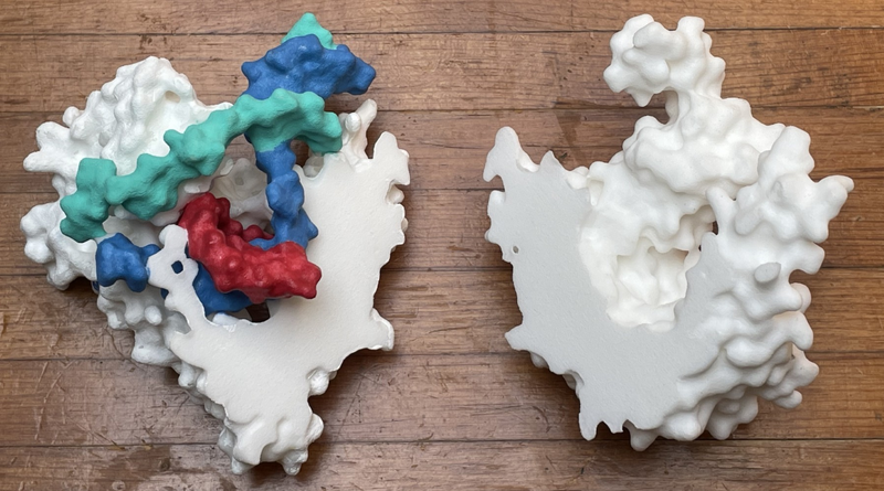
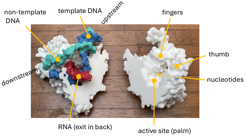
Above is a photograph of the two parts of the model. To assemble it, you bring the two flat sides together (the view is a "butterflied" version of the structure). On the left is the N-terminal part (roughly) with the nucleic acid forming a transcription bubble. On the right is the C-terminal part (roughly), in the shape of the right hand, with the active site at the palm. For an annotated image, see below.
| |||||||||||

