We apologize for Proteopedia being slow to respond. For the past two years, a new implementation of Proteopedia has been being built. Soon, it will replace this 18-year old system. All existing content will be moved to the new system at a date that will be announced here.
1w6l
From Proteopedia
(Difference between revisions)
| (14 intermediate revisions not shown.) | |||
| Line 1: | Line 1: | ||
| - | [[Image:1w6l.gif|left|200px]] | ||
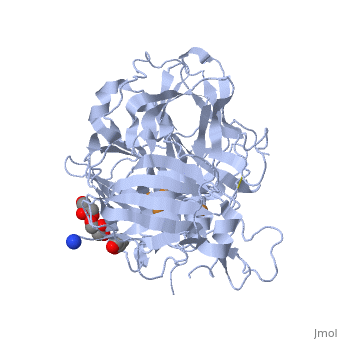
| - | < | + | ==3D structure of CotA incubated with CuCl2== |
| - | + | <StructureSection load='1w6l' size='340' side='right'caption='[[1w6l]], [[Resolution|resolution]] 2.00Å' scene=''> | |
| - | You may | + | == Structural highlights == |
| - | + | <table><tr><td colspan='2'>[[1w6l]] is a 1 chain structure with sequence from [https://en.wikipedia.org/wiki/Bacillus_subtilis Bacillus subtilis]. Full crystallographic information is available from [http://oca.weizmann.ac.il/oca-bin/ocashort?id=1W6L OCA]. For a <b>guided tour on the structure components</b> use [https://proteopedia.org/fgij/fg.htm?mol=1W6L FirstGlance]. <br> | |
| - | + | </td></tr><tr id='method'><td class="sblockLbl"><b>[[Empirical_models|Method:]]</b></td><td class="sblockDat" id="methodDat">X-ray diffraction, [[Resolution|Resolution]] 2Å</td></tr> | |
| - | + | <tr id='ligand'><td class="sblockLbl"><b>[[Ligand|Ligands:]]</b></td><td class="sblockDat" id="ligandDat"><scene name='pdbligand=CU:COPPER+(II)+ION'>CU</scene>, <scene name='pdbligand=GOL:GLYCEROL'>GOL</scene>, <scene name='pdbligand=OXY:OXYGEN+MOLECULE'>OXY</scene></td></tr> | |
| - | + | <tr id='resources'><td class="sblockLbl"><b>Resources:</b></td><td class="sblockDat"><span class='plainlinks'>[https://proteopedia.org/fgij/fg.htm?mol=1w6l FirstGlance], [http://oca.weizmann.ac.il/oca-bin/ocaids?id=1w6l OCA], [https://pdbe.org/1w6l PDBe], [https://www.rcsb.org/pdb/explore.do?structureId=1w6l RCSB], [https://www.ebi.ac.uk/pdbsum/1w6l PDBsum], [https://prosat.h-its.org/prosat/prosatexe?pdbcode=1w6l ProSAT]</span></td></tr> | |
| + | </table> | ||
| + | == Function == | ||
| + | [https://www.uniprot.org/uniprot/COTA_BACSU COTA_BACSU] Involved in brown pigmentation during sporogenesis. | ||
| + | == Evolutionary Conservation == | ||
| + | [[Image:Consurf_key_small.gif|200px|right]] | ||
| + | Check<jmol> | ||
| + | <jmolCheckbox> | ||
| + | <scriptWhenChecked>; select protein; define ~consurf_to_do selected; consurf_initial_scene = true; script "/wiki/ConSurf/w6/1w6l_consurf.spt"</scriptWhenChecked> | ||
| + | <scriptWhenUnchecked>script /wiki/extensions/Proteopedia/spt/initialview03.spt</scriptWhenUnchecked> | ||
| + | <text>to colour the structure by Evolutionary Conservation</text> | ||
| + | </jmolCheckbox> | ||
| + | </jmol>, as determined by [http://consurfdb.tau.ac.il/ ConSurfDB]. You may read the [[Conservation%2C_Evolutionary|explanation]] of the method and the full data available from [http://bental.tau.ac.il/new_ConSurfDB/main_output.php?pdb_ID=1w6l ConSurf]. | ||
| + | <div style="clear:both"></div> | ||
| + | <div style="background-color:#fffaf0;"> | ||
| + | == Publication Abstract from PubMed == | ||
| + | The multi-copper oxidases oxidise substrate molecules by accepting electrons at a mononuclear copper centre and transferring them to a trinuclear centre. Dioxygen binds to the trinuclear centre and, following the transfer of four electrons, is reduced to two molecules of water. The precise mechanism of this reduction has been unclear, but recent X-ray structural studies using the CotA endospore coat protein from Bacillus subtilis have given further insights into the principal stages. It is proposed that the mechanism involves binding of the dioxygen into the trinuclear centre so that it is sited approximately symmetrically between the two type 3 copper ions with one oxygen atom close to the type 2 copper ion. Further stages involve the formation of a peroxide intermediate and following the splitting of this intermediate, the migration of the hydroxide moieties towards the solvent exit channel. The migration steps are likely to involve a movement of the type 2 copper ion and its environment. Details of a putative mechanism are described herein based both on structures already reported in the literature and on structures of the CotA protein in the oxidised and reduced states and with the addition of peroxide and the inhibitor, azide. | ||
| - | + | Dioxygen reduction by multi-copper oxidases; a structural perspective.,Bento I, Martins LO, Gato Lopes G, Armenia Carrondo M, Lindley PF Dalton Trans. 2005 Nov 7;(21):3507-13. Epub 2005 Sep 27. PMID:16234932<ref>PMID:16234932</ref> | |
| - | + | ||
| - | + | ||
| - | + | ||
| - | + | ||
| - | + | From MEDLINE®/PubMed®, a database of the U.S. National Library of Medicine.<br> | |
| - | + | </div> | |
| + | <div class="pdbe-citations 1w6l" style="background-color:#fffaf0;"></div> | ||
| - | == | + | ==See Also== |
| - | + | *[[Laccase|Laccase]] | |
| + | *[[Laccase 3D structures|Laccase 3D structures]] | ||
| + | == References == | ||
| + | <references/> | ||
| + | __TOC__ | ||
| + | </StructureSection> | ||
[[Category: Bacillus subtilis]] | [[Category: Bacillus subtilis]] | ||
| - | [[Category: | + | [[Category: Large Structures]] |
| - | [[Category: Bento | + | [[Category: Bento I]] |
| - | [[Category: Carrondo | + | [[Category: Carrondo MA]] |
| - | [[Category: Lindley | + | [[Category: Lindley PF]] |
| - | [[Category: Lopes | + | [[Category: Lopes GG]] |
| - | [[Category: Martins | + | [[Category: Martins LO]] |
| - | + | ||
| - | + | ||
| - | + | ||
| - | + | ||
| - | + | ||
| - | + | ||
Current revision
3D structure of CotA incubated with CuCl2
| |||||||||||