This old version of Proteopedia is provided for student assignments while the new version is undergoing repairs. Content and edits done in this old version of Proteopedia after March 1, 2026 will eventually be lost when it is retired in about June of 2026.
Apply for new accounts at the new Proteopedia. Your logins will work in both the old and new versions.
1ke4
From Proteopedia
(Difference between revisions)
| (9 intermediate revisions not shown.) | |||
| Line 1: | Line 1: | ||
| - | {{Seed}} | ||
| - | [[Image:1ke4.png|left|200px]] | ||
| - | + | ==X-ray crystal structure of AmpC beta-lactamase from E. coli== | |
| - | + | <StructureSection load='1ke4' size='340' side='right'caption='[[1ke4]], [[Resolution|resolution]] 1.72Å' scene=''> | |
| - | You may | + | == Structural highlights == |
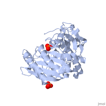
| - | + | <table><tr><td colspan='2'>[[1ke4]] is a 2 chain structure with sequence from [https://en.wikipedia.org/wiki/Escherichia_coli Escherichia coli]. Full crystallographic information is available from [http://oca.weizmann.ac.il/oca-bin/ocashort?id=1KE4 OCA]. For a <b>guided tour on the structure components</b> use [https://proteopedia.org/fgij/fg.htm?mol=1KE4 FirstGlance]. <br> | |
| - | + | </td></tr><tr id='method'><td class="sblockLbl"><b>[[Empirical_models|Method:]]</b></td><td class="sblockDat" id="methodDat">X-ray diffraction, [[Resolution|Resolution]] 1.72Å</td></tr> | |
| - | - | + | <tr id='ligand'><td class="sblockLbl"><b>[[Ligand|Ligands:]]</b></td><td class="sblockDat" id="ligandDat"><scene name='pdbligand=PO4:PHOSPHATE+ION'>PO4</scene></td></tr> |
| - | + | <tr id='resources'><td class="sblockLbl"><b>Resources:</b></td><td class="sblockDat"><span class='plainlinks'>[https://proteopedia.org/fgij/fg.htm?mol=1ke4 FirstGlance], [http://oca.weizmann.ac.il/oca-bin/ocaids?id=1ke4 OCA], [https://pdbe.org/1ke4 PDBe], [https://www.rcsb.org/pdb/explore.do?structureId=1ke4 RCSB], [https://www.ebi.ac.uk/pdbsum/1ke4 PDBsum], [https://prosat.h-its.org/prosat/prosatexe?pdbcode=1ke4 ProSAT]</span></td></tr> | |
| + | </table> | ||
| + | == Function == | ||
| + | [https://www.uniprot.org/uniprot/AMPC_ECOLI AMPC_ECOLI] This protein is a serine beta-lactamase with a substrate specificity for cephalosporins. | ||
| + | == Evolutionary Conservation == | ||
| + | [[Image:Consurf_key_small.gif|200px|right]] | ||
| + | Check<jmol> | ||
| + | <jmolCheckbox> | ||
| + | <scriptWhenChecked>; select protein; define ~consurf_to_do selected; consurf_initial_scene = true; script "/wiki/ConSurf/ke/1ke4_consurf.spt"</scriptWhenChecked> | ||
| + | <scriptWhenUnchecked>script /wiki/extensions/Proteopedia/spt/initialview01.spt</scriptWhenUnchecked> | ||
| + | <text>to colour the structure by Evolutionary Conservation</text> | ||
| + | </jmolCheckbox> | ||
| + | </jmol>, as determined by [http://consurfdb.tau.ac.il/ ConSurfDB]. You may read the [[Conservation%2C_Evolutionary|explanation]] of the method and the full data available from [http://bental.tau.ac.il/new_ConSurfDB/main_output.php?pdb_ID=1ke4 ConSurf]. | ||
| + | <div style="clear:both"></div> | ||
| + | <div style="background-color:#fffaf0;"> | ||
| + | == Publication Abstract from PubMed == | ||
| + | Beta-lactamases are the most widespread resistance mechanism to beta-lactam antibiotics and are an increasing menace to public health. Several beta-lactamase structures have been determined, making this enzyme an attractive target for structure-based drug design. To facilitate inhibitor design for the class C beta-lactamase AmpC, binding site "hot spots" on the enzyme were identified using experimental and computational approaches. Experimentally, X-ray crystal structures of AmpC in complexes with four boronic acid inhibitors and a higher resolution (1.72 A) native apo structure were determined. Along with previously determined structures of AmpC in complexes with five other boronic acid inhibitors and four beta-lactams, consensus binding sites were identified. Computationally, the programs GRID, MCSS, and X-SITE were used to predict potential binding site hot spots on AmpC. Several consensus binding sites were identified from the crystal structures. An amide recognition site was identified by the interaction between the carbonyl oxygen in the R1 side chain of beta-lactams and the atom Ndelta2 of the conserved Asn152. Surprisingly, this site also recognizes the aryl rings of arylboronic acids, appearing to form quadrupole-dipole interactions with Asn152. The highly conserved "oxyanion" hole defines a site that recognizes both carbonyl and hydroxyl groups. A hydroxyl binding site was identified by the O2 hydroxyl in the boronic acids, which hydrogen bonds with Tyr150 and a conserved water. A hydrophobic site is formed by Leu119 and Leu293. A carboxylate binding site was identified by the ubiquitous C3(4) carboxylate of the beta-lactams, which interacts with Asn346 and Arg349. Four water sites were identified by ordered waters observed in most of the structures; these waters form extensive hydrogen-bonding networks with AmpC and occasionally the ligand. Predictions by the computational programs showed some correlation with the experimentally observed binding sites. Several sites were not predicted, but novel binding sites were suggested. Taken together, a map of binding site hot spots found on AmpC, along with information on the functionality recognized at each site, was constructed. This map may be useful for structure-based inhibitor design against AmpC. | ||
| - | + | Structure-based approach for binding site identification on AmpC beta-lactamase.,Powers RA, Shoichet BK J Med Chem. 2002 Jul 18;45(15):3222-34. PMID:12109906<ref>PMID:12109906</ref> | |
| + | From MEDLINE®/PubMed®, a database of the U.S. National Library of Medicine.<br> | ||
| + | </div> | ||
| + | <div class="pdbe-citations 1ke4" style="background-color:#fffaf0;"></div> | ||
| - | + | ==See Also== | |
| - | + | *[[Beta-lactamase 3D structures|Beta-lactamase 3D structures]] | |
| - | + | == References == | |
| - | + | <references/> | |
| - | + | __TOC__ | |
| - | + | </StructureSection> | |
| - | == | + | |
| - | + | ||
| - | + | ||
| - | == | + | |
| - | + | ||
| - | + | ||
[[Category: Escherichia coli]] | [[Category: Escherichia coli]] | ||
| - | [[Category: | + | [[Category: Large Structures]] |
| - | [[Category: Powers | + | [[Category: Powers RA]] |
| - | [[Category: Shoichet | + | [[Category: Shoichet BK]] |
| - | + | ||
| - | + | ||
| - | + | ||
| - | + | ||
| - | + | ||
Current revision
X-ray crystal structure of AmpC beta-lactamase from E. coli
| |||||||||||