We apologize for Proteopedia being slow to respond. For the past two years, a new implementation of Proteopedia has been being built. Soon, it will replace this 18-year old system. All existing content will be moved to the new system at a date that will be announced here.
5dfr
From Proteopedia
(Difference between revisions)
(New page: 200px<br /><applet load="5dfr" size="450" color="white" frame="true" align="right" spinBox="true" caption="5dfr, resolution 2.3Å" /> '''CRYSTAL STRUCTURE OF ...) |
|||
| (15 intermediate revisions not shown.) | |||
| Line 1: | Line 1: | ||
| - | [[Image:5dfr.gif|left|200px]]<br /><applet load="5dfr" size="450" color="white" frame="true" align="right" spinBox="true" | ||
| - | caption="5dfr, resolution 2.3Å" /> | ||
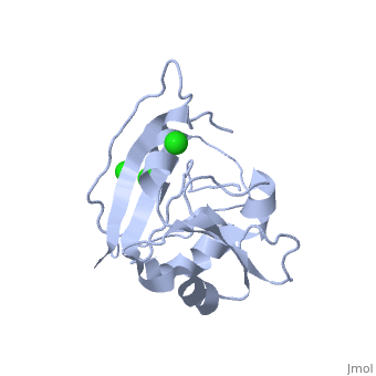
| - | '''CRYSTAL STRUCTURE OF UNLIGANDED ESCHERICHIA COLI DIHYDROFOLATE REDUCTASE. LIGAND-INDUCED CONFORMATIONAL CHANGES AND COOPERATIVITY IN BINDING'''<br /> | ||
| - | == | + | ==CRYSTAL STRUCTURE OF UNLIGANDED ESCHERICHIA COLI DIHYDROFOLATE REDUCTASE. LIGAND-INDUCED CONFORMATIONAL CHANGES AND COOPERATIVITY IN BINDING== |
| - | + | <StructureSection load='5dfr' size='340' side='right'caption='[[5dfr]], [[Resolution|resolution]] 2.30Å' scene=''> | |
| + | == Structural highlights == | ||
| + | <table><tr><td colspan='2'>[[5dfr]] is a 1 chain structure with sequence from [https://en.wikipedia.org/wiki/Escherichia_coli Escherichia coli]. Full crystallographic information is available from [http://oca.weizmann.ac.il/oca-bin/ocashort?id=5DFR OCA]. For a <b>guided tour on the structure components</b> use [https://proteopedia.org/fgij/fg.htm?mol=5DFR FirstGlance]. <br> | ||
| + | </td></tr><tr id='method'><td class="sblockLbl"><b>[[Empirical_models|Method:]]</b></td><td class="sblockDat" id="methodDat">X-ray diffraction, [[Resolution|Resolution]] 2.3Å</td></tr> | ||
| + | <tr id='ligand'><td class="sblockLbl"><b>[[Ligand|Ligands:]]</b></td><td class="sblockDat" id="ligandDat"><scene name='pdbligand=CL:CHLORIDE+ION'>CL</scene></td></tr> | ||
| + | <tr id='resources'><td class="sblockLbl"><b>Resources:</b></td><td class="sblockDat"><span class='plainlinks'>[https://proteopedia.org/fgij/fg.htm?mol=5dfr FirstGlance], [http://oca.weizmann.ac.il/oca-bin/ocaids?id=5dfr OCA], [https://pdbe.org/5dfr PDBe], [https://www.rcsb.org/pdb/explore.do?structureId=5dfr RCSB], [https://www.ebi.ac.uk/pdbsum/5dfr PDBsum], [https://prosat.h-its.org/prosat/prosatexe?pdbcode=5dfr ProSAT]</span></td></tr> | ||
| + | </table> | ||
| + | == Function == | ||
| + | [https://www.uniprot.org/uniprot/DYR_ECOLI DYR_ECOLI] Key enzyme in folate metabolism. Catalyzes an essential reaction for de novo glycine and purine synthesis, and for DNA precursor synthesis. | ||
| + | == Evolutionary Conservation == | ||
| + | [[Image:Consurf_key_small.gif|200px|right]] | ||
| + | Check<jmol> | ||
| + | <jmolCheckbox> | ||
| + | <scriptWhenChecked>; select protein; define ~consurf_to_do selected; consurf_initial_scene = true; script "/wiki/ConSurf/df/5dfr_consurf.spt"</scriptWhenChecked> | ||
| + | <scriptWhenUnchecked>script /wiki/extensions/Proteopedia/spt/initialview01.spt</scriptWhenUnchecked> | ||
| + | <text>to colour the structure by Evolutionary Conservation</text> | ||
| + | </jmolCheckbox> | ||
| + | </jmol>, as determined by [http://consurfdb.tau.ac.il/ ConSurfDB]. You may read the [[Conservation%2C_Evolutionary|explanation]] of the method and the full data available from [http://bental.tau.ac.il/new_ConSurfDB/main_output.php?pdb_ID=5dfr ConSurf]. | ||
| + | <div style="clear:both"></div> | ||
| - | == | + | ==See Also== |
| - | + | *[[Dihydrofolate reductase 3D structures|Dihydrofolate reductase 3D structures]] | |
| - | + | __TOC__ | |
| - | + | </StructureSection> | |
| - | + | ||
| - | + | ||
[[Category: Escherichia coli]] | [[Category: Escherichia coli]] | ||
| - | [[Category: | + | [[Category: Large Structures]] |
| - | [[Category: Bystroff | + | [[Category: Bystroff C]] |
| - | [[Category: Kraut | + | [[Category: Kraut J]] |
| - | + | ||
| - | + | ||
| - | + | ||
| - | + | ||
Current revision
CRYSTAL STRUCTURE OF UNLIGANDED ESCHERICHIA COLI DIHYDROFOLATE REDUCTASE. LIGAND-INDUCED CONFORMATIONAL CHANGES AND COOPERATIVITY IN BINDING
| |||||||||||

