This old version of Proteopedia is provided for student assignments while the new version is undergoing repairs. Content and edits done in this old version of Proteopedia after March 1, 2026 will eventually be lost when it is retired in about June of 2026.
Apply for new accounts at the new Proteopedia. Your logins will work in both the old and new versions.
1jfp
From Proteopedia
(Difference between revisions)
| (10 intermediate revisions not shown.) | |||
| Line 1: | Line 1: | ||
| - | {{Seed}} | ||
| - | [[Image:1jfp.png|left|200px]] | ||
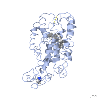
| - | < | + | ==Structure of bovine rhodopsin (dark adapted)== |
| - | + | <StructureSection load='1jfp' size='340' side='right'caption='[[1jfp]]' scene=''> | |
| - | + | == Structural highlights == | |
| - | + | <table><tr><td colspan='2'>[[1jfp]] is a 1 chain structure with sequence from [https://en.wikipedia.org/wiki/Bos_taurus Bos taurus]. Full experimental information is available from [http://oca.weizmann.ac.il/oca-bin/ocashort?id=1JFP OCA]. For a <b>guided tour on the structure components</b> use [https://proteopedia.org/fgij/fg.htm?mol=1JFP FirstGlance]. <br> | |
| - | + | </td></tr><tr id='method'><td class="sblockLbl"><b>[[Empirical_models|Method:]]</b></td><td class="sblockDat" id="methodDat">Solution NMR</td></tr> | |
| - | + | <tr id='ligand'><td class="sblockLbl"><b>[[Ligand|Ligands:]]</b></td><td class="sblockDat" id="ligandDat"><scene name='pdbligand=RET:RETINAL'>RET</scene></td></tr> | |
| - | + | <tr id='resources'><td class="sblockLbl"><b>Resources:</b></td><td class="sblockDat"><span class='plainlinks'>[https://proteopedia.org/fgij/fg.htm?mol=1jfp FirstGlance], [http://oca.weizmann.ac.il/oca-bin/ocaids?id=1jfp OCA], [https://pdbe.org/1jfp PDBe], [https://www.rcsb.org/pdb/explore.do?structureId=1jfp RCSB], [https://www.ebi.ac.uk/pdbsum/1jfp PDBsum], [https://prosat.h-its.org/prosat/prosatexe?pdbcode=1jfp ProSAT]</span></td></tr> | |
| + | </table> | ||
| + | == Function == | ||
| + | [https://www.uniprot.org/uniprot/OPSD_BOVIN OPSD_BOVIN] Photoreceptor required for image-forming vision at low light intensity. Required for photoreceptor cell viability after birth. Light-induced isomerization of 11-cis to all-trans retinal triggers a conformational change leading to G-protein activation and release of all-trans retinal (By similarity).<ref>PMID:16908857</ref> <ref>PMID:17060607</ref> | ||
| + | == Evolutionary Conservation == | ||
| + | [[Image:Consurf_key_small.gif|200px|right]] | ||
| + | Check<jmol> | ||
| + | <jmolCheckbox> | ||
| + | <scriptWhenChecked>; select protein; define ~consurf_to_do selected; consurf_initial_scene = true; script "/wiki/ConSurf/jf/1jfp_consurf.spt"</scriptWhenChecked> | ||
| + | <scriptWhenUnchecked>script /wiki/extensions/Proteopedia/spt/initialview01.spt</scriptWhenUnchecked> | ||
| + | <text>to colour the structure by Evolutionary Conservation</text> | ||
| + | </jmolCheckbox> | ||
| + | </jmol>, as determined by [http://consurfdb.tau.ac.il/ ConSurfDB]. You may read the [[Conservation%2C_Evolutionary|explanation]] of the method and the full data available from [http://bental.tau.ac.il/new_ConSurfDB/main_output.php?pdb_ID=1jfp ConSurf]. | ||
| + | <div style="clear:both"></div> | ||
| - | == | + | ==See Also== |
| - | + | *[[Rhodopsin|Rhodopsin]] | |
| - | + | *[[Rhodopsin 3D structures|Rhodopsin 3D structures]] | |
| - | + | == References == | |
| - | + | <references/> | |
| - | + | __TOC__ | |
| - | + | </StructureSection> | |
| - | + | ||
| - | + | ||
| - | + | ||
| - | + | ||
| - | + | ||
| - | == | + | |
| - | < | + | |
[[Category: Bos taurus]] | [[Category: Bos taurus]] | ||
| - | [[Category: | + | [[Category: Large Structures]] |
| - | [[Category: | + | [[Category: Albert AD]] |
| - | [[Category: | + | [[Category: Choi G]] |
| - | [[Category: | + | [[Category: Yeagle PL]] |
| - | + | ||
| - | + | ||
Current revision
Structure of bovine rhodopsin (dark adapted)
| |||||||||||