This old version of Proteopedia is provided for student assignments while the new version is undergoing repairs. Content and edits done in this old version of Proteopedia after March 1, 2026 will eventually be lost when it is retired in about June of 2026.
Apply for new accounts at the new Proteopedia. Your logins will work in both the old and new versions.
2ghj
From Proteopedia
(Difference between revisions)
| (7 intermediate revisions not shown.) | |||
| Line 1: | Line 1: | ||
| - | {{Seed}} | ||
| - | [[Image:2ghj.png|left|200px]] | ||
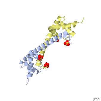
| - | + | ==Crystal structure of folded and partially unfolded forms of Aquifex aeolicus ribosomal protein L20== | |
| - | + | <StructureSection load='2ghj' size='340' side='right'caption='[[2ghj]], [[Resolution|resolution]] 2.90Å' scene=''> | |
| - | + | == Structural highlights == | |
| - | + | <table><tr><td colspan='2'>[[2ghj]] is a 4 chain structure with sequence from [https://en.wikipedia.org/wiki/"aquifex_aeolicus"_huber_and_stetter_2001 "aquifex aeolicus" huber and stetter 2001]. Full crystallographic information is available from [http://oca.weizmann.ac.il/oca-bin/ocashort?id=2GHJ OCA]. For a <b>guided tour on the structure components</b> use [https://proteopedia.org/fgij/fg.htm?mol=2GHJ FirstGlance]. <br> | |
| - | + | </td></tr><tr id='ligand'><td class="sblockLbl"><b>[[Ligand|Ligands:]]</b></td><td class="sblockDat" id="ligandDat"><scene name='pdbligand=SO4:SULFATE+ION'>SO4</scene></td></tr> | |
| - | - | + | <tr id='NonStdRes'><td class="sblockLbl"><b>[[Non-Standard_Residue|NonStd Res:]]</b></td><td class="sblockDat"><scene name='pdbligand=MSE:SELENOMETHIONINE'>MSE</scene></td></tr> |
| - | + | <tr id='gene'><td class="sblockLbl"><b>[[Gene|Gene:]]</b></td><td class="sblockDat">rplT ([https://www.ncbi.nlm.nih.gov/Taxonomy/Browser/wwwtax.cgi?mode=Info&srchmode=5&id=63363 "Aquifex aeolicus" Huber and Stetter 2001])</td></tr> | |
| + | <tr id='resources'><td class="sblockLbl"><b>Resources:</b></td><td class="sblockDat"><span class='plainlinks'>[https://proteopedia.org/fgij/fg.htm?mol=2ghj FirstGlance], [http://oca.weizmann.ac.il/oca-bin/ocaids?id=2ghj OCA], [https://pdbe.org/2ghj PDBe], [https://www.rcsb.org/pdb/explore.do?structureId=2ghj RCSB], [https://www.ebi.ac.uk/pdbsum/2ghj PDBsum], [https://prosat.h-its.org/prosat/prosatexe?pdbcode=2ghj ProSAT]</span></td></tr> | ||
| + | </table> | ||
| + | == Function == | ||
| + | [[https://www.uniprot.org/uniprot/RL20_AQUAE RL20_AQUAE]] Binds directly to 23S ribosomal RNA and is necessary for the in vitro assembly process of the 50S ribosomal subunit. It is not involved in the protein synthesizing functions of that subunit (By similarity). | ||
| + | == Evolutionary Conservation == | ||
| + | [[Image:Consurf_key_small.gif|200px|right]] | ||
| + | Check<jmol> | ||
| + | <jmolCheckbox> | ||
| + | <scriptWhenChecked>; select protein; define ~consurf_to_do selected; consurf_initial_scene = true; script "/wiki/ConSurf/gh/2ghj_consurf.spt"</scriptWhenChecked> | ||
| + | <scriptWhenUnchecked>script /wiki/extensions/Proteopedia/spt/initialview01.spt</scriptWhenUnchecked> | ||
| + | <text>to colour the structure by Evolutionary Conservation</text> | ||
| + | </jmolCheckbox> | ||
| + | </jmol>, as determined by [http://consurfdb.tau.ac.il/ ConSurfDB]. You may read the [[Conservation%2C_Evolutionary|explanation]] of the method and the full data available from [http://bental.tau.ac.il/new_ConSurfDB/main_output.php?pdb_ID=2ghj ConSurf]. | ||
| + | <div style="clear:both"></div> | ||
| + | <div style="background-color:#fffaf0;"> | ||
| + | == Publication Abstract from PubMed == | ||
| + | The recent finding of intrinsically unstructured proteins defies the classical structure-function paradigm. However, owing to their flexibility, intrinsically unstructured proteins generally escape detailed structural investigations. Consequently little is known about the extent of conformational disorder and its role in biological functions. Here, we present the X-ray structure of the unbound ribosomal protein L20, the long basic amino-terminal extension of which has been previously interpreted as fully disordered in the absence of RNA. This study provides the first detailed picture of two protein folding states trapped together in a crystal and indicates that unfolding occurs in discrete regions of the whole protein, corresponding mainly to RNA-binding residues. The electrostatic destabilization of the long alpha-helix and a structural communication between the two L20 domains are reminiscent of those observed in calmodulin. The detailed comparison of the two conformations observed in the crystal provides new insights into the role of unfolded extensions in ribosomal assembly. | ||
| - | + | Coexistence of two protein folding states in the crystal structure of ribosomal protein L20.,Timsit Y, Allemand F, Chiaruttini C, Springer M EMBO Rep. 2006 Oct;7(10):1013-8. Epub 2006 Sep 15. PMID:16977336<ref>PMID:16977336</ref> | |
| + | From MEDLINE®/PubMed®, a database of the U.S. National Library of Medicine.<br> | ||
| + | </div> | ||
| + | <div class="pdbe-citations 2ghj" style="background-color:#fffaf0;"></div> | ||
| - | + | ==See Also== | |
| - | + | *[[Ribosomal protein L20|Ribosomal protein L20]] | |
| - | + | == References == | |
| - | + | <references/> | |
| - | + | __TOC__ | |
| - | + | </StructureSection> | |
| - | == | + | [[Category: Aquifex aeolicus huber and stetter 2001]] |
| - | + | [[Category: Large Structures]] | |
| - | + | [[Category: Allemand, F]] | |
| - | == | + | [[Category: Chiaruttini, C]] |
| - | < | + | [[Category: Springer, M]] |
| - | [[Category: Aquifex aeolicus]] | + | [[Category: Timsit, Y]] |
| - | [[Category: Allemand, F | + | |
| - | [[Category: Chiaruttini, C | + | |
| - | [[Category: Springer, M | + | |
| - | [[Category: Timsit, Y | + | |
[[Category: Folding intermediate]] | [[Category: Folding intermediate]] | ||
[[Category: Ribosomal protein extension]] | [[Category: Ribosomal protein extension]] | ||
| - | + | [[Category: Structural protein]] | |
| - | + | ||
Current revision
Crystal structure of folded and partially unfolded forms of Aquifex aeolicus ribosomal protein L20
| |||||||||||