This old version of Proteopedia is provided for student assignments while the new version is undergoing repairs. Content and edits done in this old version of Proteopedia after March 1, 2026 will eventually be lost when it is retired in about June of 2026.
Apply for new accounts at the new Proteopedia. Your logins will work in both the old and new versions.
3hay
From Proteopedia
(Difference between revisions)
(New page: '''Unreleased structure''' The entry 3hay is ON HOLD Authors: Ye, K. Description: Crystal structure of a substrate-bound full H/ACA RNP from Pyrococcus furiosus ''Page seeded by [http...) |
|||
| (11 intermediate revisions not shown.) | |||
| Line 1: | Line 1: | ||
| - | '''Unreleased structure''' | ||
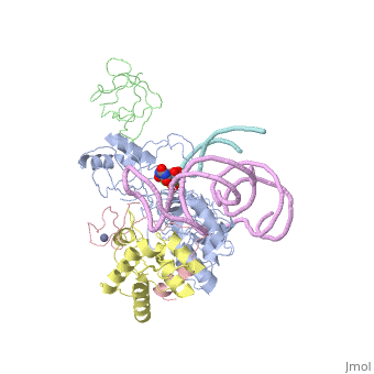
| - | + | ==Crystal structure of a substrate-bound full H/ACA RNP from Pyrococcus furiosus== | |
| + | <StructureSection load='3hay' size='340' side='right'caption='[[3hay]], [[Resolution|resolution]] 4.99Å' scene=''> | ||
| + | == Structural highlights == | ||
| + | <table><tr><td colspan='2'>[[3hay]] is a 6 chain structure with sequence from [https://en.wikipedia.org/wiki/Pyrococcus_furiosus Pyrococcus furiosus]. Full crystallographic information is available from [http://oca.weizmann.ac.il/oca-bin/ocashort?id=3HAY OCA]. For a <b>guided tour on the structure components</b> use [https://proteopedia.org/fgij/fg.htm?mol=3HAY FirstGlance]. <br> | ||
| + | </td></tr><tr id='method'><td class="sblockLbl"><b>[[Empirical_models|Method:]]</b></td><td class="sblockDat" id="methodDat">X-ray diffraction, [[Resolution|Resolution]] 4.99Å</td></tr> | ||
| + | <tr id='ligand'><td class="sblockLbl"><b>[[Ligand|Ligands:]]</b></td><td class="sblockDat" id="ligandDat"><scene name='pdbligand=FHU:(5S,6R)-5-FLUORO-6-HYDROXY-PSEUDOURIDINE-5-MONOPHOSPHATE'>FHU</scene>, <scene name='pdbligand=ZN:ZINC+ION'>ZN</scene></td></tr> | ||
| + | <tr id='resources'><td class="sblockLbl"><b>Resources:</b></td><td class="sblockDat"><span class='plainlinks'>[https://proteopedia.org/fgij/fg.htm?mol=3hay FirstGlance], [http://oca.weizmann.ac.il/oca-bin/ocaids?id=3hay OCA], [https://pdbe.org/3hay PDBe], [https://www.rcsb.org/pdb/explore.do?structureId=3hay RCSB], [https://www.ebi.ac.uk/pdbsum/3hay PDBsum], [https://prosat.h-its.org/prosat/prosatexe?pdbcode=3hay ProSAT]</span></td></tr> | ||
| + | </table> | ||
| + | == Function == | ||
| + | [https://www.uniprot.org/uniprot/TRUB_PYRFU TRUB_PYRFU] Could be responsible for synthesis of pseudouridine from uracil-55 in the psi GC loop of transfer RNAs (By similarity). | ||
| + | == Evolutionary Conservation == | ||
| + | [[Image:Consurf_key_small.gif|200px|right]] | ||
| + | Check<jmol> | ||
| + | <jmolCheckbox> | ||
| + | <scriptWhenChecked>; select protein; define ~consurf_to_do selected; consurf_initial_scene = true; script "/wiki/ConSurf/ha/3hay_consurf.spt"</scriptWhenChecked> | ||
| + | <scriptWhenUnchecked>script /wiki/extensions/Proteopedia/spt/initialview01.spt</scriptWhenUnchecked> | ||
| + | <text>to colour the structure by Evolutionary Conservation</text> | ||
| + | </jmolCheckbox> | ||
| + | </jmol>, as determined by [http://consurfdb.tau.ac.il/ ConSurfDB]. You may read the [[Conservation%2C_Evolutionary|explanation]] of the method and the full data available from [http://bental.tau.ac.il/new_ConSurfDB/main_output.php?pdb_ID=3hay ConSurf]. | ||
| + | <div style="clear:both"></div> | ||
| + | <div style="background-color:#fffaf0;"> | ||
| + | == Publication Abstract from PubMed == | ||
| + | H/ACA RNAs form ribonucleoprotein complex (RNP) with proteins Cbf5, Nop10, L7Ae, and Gar1 and guide site-specific conversion of uridine into pseudouridine in cellular RNAs. The crystal structures of H/ACA RNP with substrate bound at the active site cleft reveal that the substrate is recruited through sequence-specific pairing with guide RNA and essential protein contacts. Substrate binding leads to a reorganization of a preset pseudouridylation pocket and an adaptive movement of the PUA domain and the lower stem of the H/ACA RNA. Moreover, a thumb loop flips from the Gar1-bound state in the substrate-free RNP structure to tightly associate with the substrate. Mutagenesis and enzyme kinetics analysis suggest a critical role of Gar1 and the thumb in substrate turnover, particularly in product release. Comparison with tRNA Psi55 synthase TruB reveals the structural conservation and adaptation between an RNA-guided and stand-alone pseudouridine synthase and provides insight into the guide-independent activity of Cbf5. | ||
| - | + | Structural mechanism of substrate RNA recruitment in H/ACA RNA-guided pseudouridine synthase.,Duan J, Li L, Lu J, Wang W, Ye K Mol Cell. 2009 May 14;34(4):427-39. PMID:19481523<ref>PMID:19481523</ref> | |
| - | + | From MEDLINE®/PubMed®, a database of the U.S. National Library of Medicine.<br> | |
| + | </div> | ||
| + | <div class="pdbe-citations 3hay" style="background-color:#fffaf0;"></div> | ||
| - | + | ==See Also== | |
| + | *[[Guide-independent Pseudouridine synthase|Guide-independent Pseudouridine synthase]] | ||
| + | *[[Kink-turn motif|Kink-turn motif]] | ||
| + | *[[Pseudouridine synthase 3D structures|Pseudouridine synthase 3D structures]] | ||
| + | *[[Ribosome biogenesis protein|Ribosome biogenesis protein]] | ||
| + | *[[Ribosome biogenesis protein 3D structures|Ribosome biogenesis protein 3D structures]] | ||
| + | *[[User:Wayne Decatur/kink-turn motif|User:Wayne Decatur/kink-turn motif]] | ||
| + | == References == | ||
| + | <references/> | ||
| + | __TOC__ | ||
| + | </StructureSection> | ||
| + | [[Category: Large Structures]] | ||
| + | [[Category: Pyrococcus furiosus]] | ||
| + | [[Category: Ye K]] | ||
Current revision
Crystal structure of a substrate-bound full H/ACA RNP from Pyrococcus furiosus
| |||||||||||