From Proteopedia
< User:Gisselle Medina(Difference between revisions)
proteopedia linkproteopedia link
|
|
| (11 intermediate revisions not shown.) |
| Line 1: |
Line 1: |
| - | <applet load='1U19' size='300' frame='true' align='right' caption='Rhodopsin' /> | + | <applet load='1U19' size='200' frame='true' align='right' caption='Rhodopsin' /> |
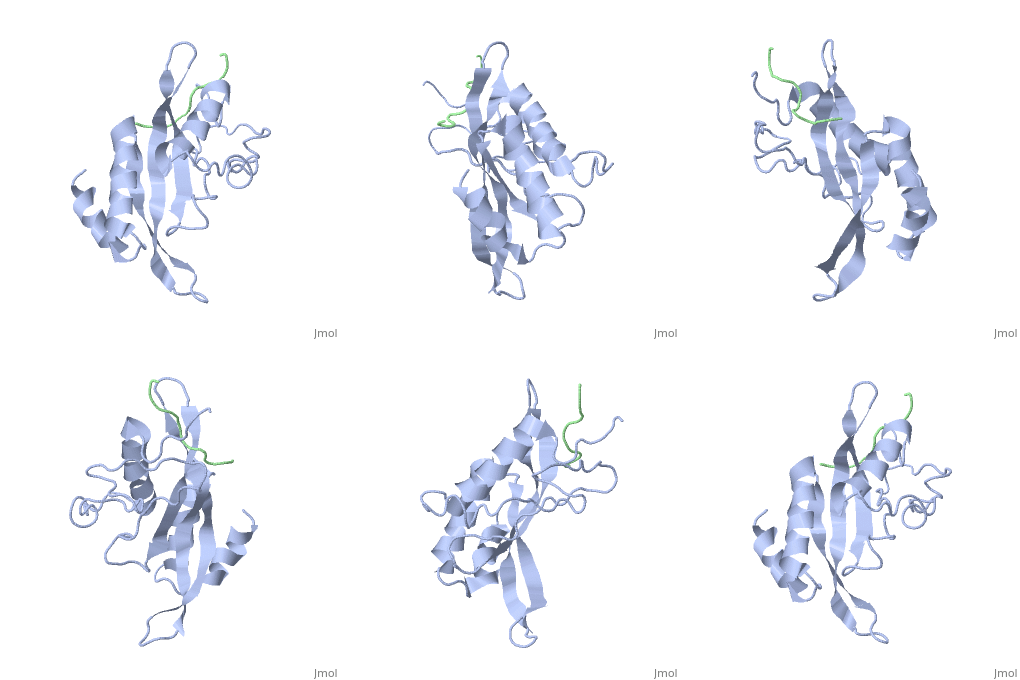
| | + | <applet load='3CZB' size='200' frame='true' align='right' caption='Carbonic Anhydrase' /> |
| | + | <applet load='1lqb' size='200' frame='true' align='right' caption='HIF-1 alpha' /> |
| | + | <applet load='1m4p' size='200' frame='true' align='right' caption='Tsg101-PTAP' /> |
| | | | |
| - | == Abstract == | + | <scene name='User:Gisselle_Medina/Sandbox2_Rhodopsin/Ptap/4'>Tsg101-PTAP</scene> |
| - | The structure of the active state of rhodopsin has only been isolated recently. We have highlighted certain amino acids in the RasMol model to demonstrate rhodopsin's activated state, Meta II (Figure 1). The extracellular loop two (EL2) in blue, changes conformation in what has been described as a hinge-like motion (Ahuja et. al 2009). Met207 (purple) located in the inner helix five (H5), stabilizes the β-ionone ring in the chromophore, specifically the C5/C6/C7/C8 region during the inactivated state (not shown). The amino acids Cys167 (green) and His211 (red) are also in close proximity to the chromophore and aid in the stabilization of the retinal during the inactivation. Met207 and His211 maintain their interactions with the retinal during Meta II. There is a loss of interaction between Cys167 and the retinal and as well as between Cys167 and His211 during this state. However, Met207 interacts with Cys167 which supports the idea that H5 loop changes conformation in concert with the movement EL2. (Ahuja et. al 2008)
| + | |
Current revision

