This old version of Proteopedia is provided for student assignments while the new version is undergoing repairs. Content and edits done in this old version of Proteopedia after March 1, 2026 will eventually be lost when it is retired in about June of 2026.
Apply for new accounts at the new Proteopedia. Your logins will work in both the old and new versions.
User:Eleanor Crabb/Sandbox 2
From Proteopedia
| Line 1: | Line 1: | ||
| - | + | '''The four tiers of protein structure''' | |
| - | + | We will use the structure of the zinc-containing enzyme '''1z3a''' to identify the four tiers of protein structure. <applet load='1z3a' size='300' frame='true' align='right' caption='Insert caption here' /> | |
| - | + | ||
| - | The | + | The '''primary structure''' of a protein is simply the arrangement of amino acid residues within the chains. This can be emphasized by colouring just the chains according to <scene name='User:Eleanor_Crabb/Sandbox_2/1z3a_chaina_amino/1'>residue</scene> (in this example we will use just Chain A to illustrate this). Hover your cursor over the molecule to identify some of the different residues present. |
| - | + | The protein can also be coloured according to '''secondary structure'''. The elements of <scene name='User:Eleanor_Crabb/Sandbox_2/1z3a_chaina_secondarystructure/1'>secondary structure</scene> in this protein are the alpha-helices shown in pink and the beta-sheets in yellow. The interconnecting loops and any other features are shown in white. Rotate the molecule to look at these elements in more detail. If we now consider just one <scene name='User:Eleanor_Crabb/Sandbox_2/1z3a_ahelix_hbonds/1'>alpha-helix</scene> we can see how this secondary structure is held together by hydrogen bonds - shown here as dashed lines. Remember that the H atoms are not displayed in the protein so the lines join the atoms that are hydrogen bonded together (without the bonded hydrogen atom). The <scene name='User:Eleanor_Crabb/Sandbox_2/1z3a_bsheet_hydrogenbnds/1'>hydrogen bonding in the beta-sheet</scene> can also be illustrated. | |
| - | Finally, the quaternary structure | + | As an enzyme, this protein is an example of a globular protein as we can see when consider the <scene name='User:Eleanor_Crabb/Sandbox_2/1z3a_spacefilled/1'>space-filled representation</scene> of the entire protein, both chains A and B. This the '''tertiary structure''' of the protein. |
| + | |||
| + | Finally, the '''quaternary structure''' refers to the number of subunits in the protein. In this particular example, the biological molecule is the same as the shown <scene name='User:Eleanor_Crabb/Sandbox_2/1z3a_chainaandb/1'>asymmetric unit of 1z3a</scene> shown here and so there are two. The protein is thus a dimer. | ||
Revision as of 14:59, 20 October 2009
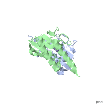
The four tiers of protein structure
We will use the structure of the zinc-containing enzyme 1z3a to identify the four tiers of protein structure.
|
The primary structure of a protein is simply the arrangement of amino acid residues within the chains. This can be emphasized by colouring just the chains according to (in this example we will use just Chain A to illustrate this). Hover your cursor over the molecule to identify some of the different residues present.
The protein can also be coloured according to secondary structure. The elements of in this protein are the alpha-helices shown in pink and the beta-sheets in yellow. The interconnecting loops and any other features are shown in white. Rotate the molecule to look at these elements in more detail. If we now consider just one we can see how this secondary structure is held together by hydrogen bonds - shown here as dashed lines. Remember that the H atoms are not displayed in the protein so the lines join the atoms that are hydrogen bonded together (without the bonded hydrogen atom). The can also be illustrated.
As an enzyme, this protein is an example of a globular protein as we can see when consider the of the entire protein, both chains A and B. This the tertiary structure of the protein.
Finally, the quaternary structure refers to the number of subunits in the protein. In this particular example, the biological molecule is the same as the shown shown here and so there are two. The protein is thus a dimer.
