|
|
| Line 1: |
Line 1: |
| - | __NOTOC__
| + | {{Seed}} |
| - | {{Quote box | + | [[Image:1xbn.png|left|200px]] |
| - | | quote = <b>Hemoglobin causes Net Diffusion of Oxygen</b><br>Oxygen diffuses freely across oxygen-permeable membranes, following the simple rule of equal pressure. By capturing oxygen, hemoglobin produces a change in the internal pressure of free oxygen, forcing the oxygen influx where it is needed.
| + | |
| - | | source = [http://highered.mcgraw-hill.com/olcweb/cgi/pluginpop.cgi?it=swf::640::480::/sites/dl/free/0077290828/811360/Hemoglobin_Causes_Net_Diffusion_of_Oxygen.swf::Hemoglobin%20Causes%20Net%20Diffusion%20of%20Oxygen (interactive demo)]
| + | |
| - | | width = 40%
| + | |
| - | | align = right
| + | |
| - | }}
| + | |
| | | | |
| - | ===How do we get the oxygen we breathe?===
| + | <!-- |
| - | Oxygen is required by all aerobic animals as a basic mechanism to accept electrons and hydrogen ions produced during metabolism. Even when oxygen is all around us, in the air and in the water, oxygen need to be close to the cells where it's going to be used.
| + | The line below this paragraph, containing "STRUCTURE_1xbn", creates the "Structure Box" on the page. |
| | + | You may change the PDB parameter (which sets the PDB file loaded into the applet) |
| | + | or the SCENE parameter (which sets the initial scene displayed when the page is loaded), |
| | + | or leave the SCENE parameter empty for the default display. |
| | + | --> |
| | + | {{STRUCTURE_1xbn| PDB=1xbn | SCENE= }} |
| | | | |
| - | This is the time when blood comes into action. Blood is a specialized body fluid that delivers necessary substances to the body's cells – such as nutrients and oxygen – and transports waste products away from those same cells. Because oxygen is not very soluble in blood, animals have an efficient mechanism for capturing oxygen, transporting, and releasing it by the inner cells: hemoglobin.
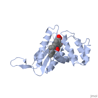
| + | ===Crystal structure of a bacterial nitric oxide sensor: an ortholog of mammalian soluble guanylate cyclase heme domain=== |
| - | {{Clear}}
| + | |
| - | ===Hb, the mighty protein=== | + | |
| - | Hemoglobin (Hb), also spelled haemoglobin, (see on the right a three-dimensional representation of a single molecule) is the protein that carries oxygen from the lungs to the tissues where it's needed. The carbon dioxide produced by the consumption of oxygen. In order to function most efficiently, hemoglobin needs to bind to oxygen tightly in the oxygen-rich atmosphere of the lungs and be able to release oxygen rapidly in the relatively oxygen-poor environment of the tissues. It does this in a most elegant and intricately coordinated way. <u>The story of hemoglobin is the prototype example of the relationship between structure and function of a protein molecule</u>.
| + | |
| - | <applet load='' size='300' frame='true' align='right' caption='' scene='User:Jaime_Prilusky/How_do_we_get_the_oxygen_we_breathe/1hho_bio/1'/>
| + | |
| | | | |
| - | =====The true colors===== | |
| - | As you can see, there are <scene name='User:Jaime_Prilusky/How_do_we_get_the_oxygen_we_breathe/1hho_bio/4'>four</scene> identical gray, red, blue and orange colored, made-of-balls elements. Let's take a closer look to <scene name='User:Jaime_Prilusky/How_do_we_get_the_oxygen_we_breathe/1hho_bio/6'>one</scene> of them. This is the ''''heme'''' group, the functional unit of hemoglobin. | |
| - | ''WAIT'': are those the true colors of the heme group? Not really. We are looking at a representation of the real structure, artificially colored following the [[CPK|Corey-Pauling-Koltun]] scheme ( {{Template:ColorKey_Element_C}} | |
| - | {{Template:ColorKey_Element_H}} | |
| - | {{Template:ColorKey_Element_O}} | |
| - | {{Template:ColorKey_Element_N}} | |
| - | {{Template:ColorKey_Element_S}} | |
| - | {{Template:ColorKey_Element_Fe}} ). ''Remember'': these are artificial representations, using colors, textures, styles and forms chosen with the purpose of helping us to better understand the reality in its rich spacial configuration. | |
| - | This model of the heme group is represented here in a | |
| - | <scene name='User:Jaime_Prilusky/How_do_we_get_the_oxygen_we_breathe/1hho_hem_styles/1'>spacefill</scene> mode, but we can also draw it as | |
| - | <scene name='User:Jaime_Prilusky/How_do_we_get_the_oxygen_we_breathe/1hho_hem_styles/2'>ball and sticks</scene>, maintaining always the same spacial structure and color coded information. | |
| | | | |
| - | =====Capturing Oxygen and other molecules ...=====
| + | <!-- |
| - | The "heart" of the hemoglobin is the <scene name='User:Jaime_Prilusky/How_do_we_get_the_oxygen_we_breathe/Heme_deoxy/2'>heme</scene> group which is a flat ring molecule containing {{Template:ColorKey_Element_C}}arbon, {{Template:ColorKey_Element_N}}itrogen and {{Template:ColorKey_Element_H}}ydrogen atoms, with a single <font color="#E06633">Fe2+</font> ion at the center. In a heme molecule, the iron is held within the flat plane by four nitrogen ligands from that ring (rotate the structure with your mouse to see the flat plane from its side). <!-- The iron ion makes a fifth bond to a histidine side chain from one of polypeptide chain that forms the heme pocket. --> In the proper conditions, an oxygen molecule gets | + | The line below this paragraph, {{ABSTRACT_PUBMED_15472039}}, adds the Publication Abstract to the page |
| - | <scene name='User:Jaime_Prilusky/How_do_we_get_the_oxygen_we_breathe/Heme/1'>attached to the Fe</scene> in the heme group. ''OBSERVE'' Are there other changes besides the oxygen being attached to the Fe?
| + | (as it appears on PubMed at http://www.pubmed.gov), where 15472039 is the PubMed ID number. |
| - | We can watch the capturing of an oxygen molecule in the context of a <scene name='User:Jaime_Prilusky/How_do_we_get_the_oxygen_we_breathe/Heme/2'>protein single chain</scene> or on a close-up view of the <scene name='User:Jaime_Prilusky/How_do_we_get_the_oxygen_we_breathe/Heme/1'>isolated Heme</scene> group. {{Template:Button Toggle Animation2}}
| + | --> |
| | + | {{ABSTRACT_PUBMED_15472039}} |
| | | | |
| - | And now is when things get interesting. The hem group has the chemical and structural capabilities to capture an <scene name='User:Jaime_Prilusky/How_do_we_get_the_oxygen_we_breathe/O2/1'>oxygen</scene> molecule, which happens to be too close to the general shape of a molecule of <scene name='User:Jaime_Prilusky/How_do_we_get_the_oxygen_we_breathe/Co/1'>carbon monoxide</scene>, which binds hemoglobin about 240 times faster and better than oxygen, meaning that if both gases are available, hemoglobin will prefer CO over O2. ''THINK'': Can you imagine what will happen if by accident we breathe in a carbon monoxide rich atmosphere?
| + | ==About this Structure== |
| | + | 1XBN is a 1 chain structure of sequence from [http://en.wikipedia.org/wiki/Thermoanaerobacter_tengcongensis Thermoanaerobacter tengcongensis]. Full crystallographic information is available from [http://oca.weizmann.ac.il/oca-bin/ocashort?id=1XBN OCA]. |
| | | | |
| - | =====The whole molecule===== | + | ==Reference== |
| - | Let's go back and take a look to the whole picture. Do you remember the four '''heme''' groups in a ribbon-like structure we noticed at the beginning?. This is because the biological active molecule of hemoglobin is a ''tetramer'', this is, a polymer comprising four monomer units: two alpha chains, each with 141 amino acids and two beta chains, each with 146 amino acids. The protein of each of these chains is called ''globin''.
| + | <ref group="xtra">PMID:15472039</ref><references group="xtra"/> |
| | + | [[Category: Thermoanaerobacter tengcongensis]] |
| | + | [[Category: Nioche, P.]] |
| | + | [[Category: Raman, C S.]] |
| | + | [[Category: Cgmp]] |
| | + | [[Category: Heme protein]] |
| | + | [[Category: Nitric oxide]] |
| | + | [[Category: Soluble guanylyl cyclase]] |
| | | | |
| - | ===Sickle-cell disease===
| + | ''Page seeded by [http://oca.weizmann.ac.il/oca OCA ] on Tue Feb 17 06:48:42 2009'' |
| - | Sickle hemoglobin differs from normal hemoglobin by a single amino acid: valine (hydrophobic) replaces glutamate (hydrophilic) at position 6 on the surface of the beta chain. This creates an hydrophobic spot. ''THINK'': Why a simple additional hydrophobic spot (actually two spots in the structure ''WHY?''), generated by the change of a single amino acid on a protein with over 500 amino acids becomes so problematic?
| + | |
| - | <applet load='1hbs' size='300' frame='true' align='right' caption='' scene='User:Jaime_Prilusky/How_do_we_get_the_oxygen_we_breathe/Sickle_one_protein/4'/>
| + | |
| - | On the right, we can see the structure of a deoxygenated hemoglobin, this is, an hemoglobin shortly after releasing the load of oxygen. We can distinguish it's four chains (by it's artificial colors) and the four heme groups with no oxygen attached. This time, the representation is of style ''spacefill'', which is Ok because you know by now that representations are only a different way of drawing a real structure that we can't see.
| + | |
| - | | + | |
| - | Both normal and sickle hemoglobin, when in deoxygenated state, have an
| + | |
| - | <scene name='User:Jaime_Prilusky/How_do_we_get_the_oxygen_we_breathe/Dexygenated_hemoglobin/3'>hydrophobic spot</scene> (colored white here)
| + | |
| - | on the beta chains. Two beta chains = two hydrophobic spots on the dehydrogenated hemoglobin. ''WATCH'': Can you find the spots on the two chains?. | + | |
| - | | + | |
| - | The <scene name='User:Jaime_Prilusky/How_do_we_get_the_oxygen_we_breathe/Sickle_hemoglobin/1'>hydrophobic spot</scene> present on Sickle hemoglobin sticks to the hydrophobic spot present on dehydrogenized hemoglobin, causing hemoglobin molecules to
| + | |
| - | <scene name='User:Jaime_Prilusky/How_do_we_get_the_oxygen_we_breathe/Sickle_hemoglobin_chain/1'>aggregate</scene> into chains forming long fibers.
| + | |
| - | A <scene name='User:Jaime_Prilusky/How_do_we_get_the_oxygen_we_breathe/Sickle_hemoglobin_chain_close/1'>closer look</scene>
| + | |
| - | shows us that Alanine and Leucine from one molecule attract the Valine from another, chaining the two hemoglobin molecules together.
| + | |
| - | | + | |
| - | {{Clear}}
| + | |
| - | ==Content advisors==
| + | |
| - | This lesson plan was developed together with Dr. Dvora Cohen, Biology Teacher and Dr Mira Kipnis, Chemistry Teacher, both from the Davidson Institute of Science Education, Weizmann Institute of Science. This page include scenes, structures and ideas from [[User:Eric_Martz|Eric Martz]], [[User:Frieda S. Reichsman|Frieda S. Reichsman]] and [[User:Angel_Herraez|Angel Herraez]].
| + | |

