Colicin E1
From Proteopedia
| Line 23: | Line 23: | ||
The cytocoxic domain of Colicin R1 forms a channel in the membrane of the ''E. coli'' cell once it has passed into the cell. The opening of the channel is controlled in a lipid-dependent manner by a histadine residue at 440. The open channel changes the rate of increase in the membrane conductance, and this change is maximum at an acidic pH. Once bound, there is a pH shift from 4 to 6 on both sides of the membrane. This causes a large increase in the trans-membrane current. | The cytocoxic domain of Colicin R1 forms a channel in the membrane of the ''E. coli'' cell once it has passed into the cell. The opening of the channel is controlled in a lipid-dependent manner by a histadine residue at 440. The open channel changes the rate of increase in the membrane conductance, and this change is maximum at an acidic pH. Once bound, there is a pH shift from 4 to 6 on both sides of the membrane. This causes a large increase in the trans-membrane current. | ||
| - | Alkalinization-induced weakening of the electrostatic interactions between colicin and the membrane surface facilitates conformational changes required for the transition of membrane-bound colicin molecules to an active channel state. | + | Alkalinization-induced weakening of the electrostatic interactions between colicin and the membrane surface facilitates conformational changes required for the transition of membrane-bound colicin molecules to an active channel state. The channel activity of colicin E1 is not monotonically dependent on the magnitude of the negative surface potential of the target membrane. |
Introducing a His440Ala mutation (changing a positive residue to a neutral one) eliminates the pH-shift effect, showing that the change is associated with deprotonation of the His residue, which occurs at an acidic pH. An H427A mutation behaves similarly to the wild type, indicating that it is the deprotonisation of His440 that induces the alkalinization activation. His 440 is located near to the lipid head groups of the bilayer, and His 427 is in the helix that is translocated to the trans-side of the membrane when the channel is open. | Introducing a His440Ala mutation (changing a positive residue to a neutral one) eliminates the pH-shift effect, showing that the change is associated with deprotonation of the His residue, which occurs at an acidic pH. An H427A mutation behaves similarly to the wild type, indicating that it is the deprotonisation of His440 that induces the alkalinization activation. His 440 is located near to the lipid head groups of the bilayer, and His 427 is in the helix that is translocated to the trans-side of the membrane when the channel is open. | ||
| + | |||
| + | [[Image:From 28.png]] | ||
The in vitro activity of channel-forming colicins is largest at an acidic ambient pH - colicin E1 is maximum at less than pH5, with a membrane potential of -60mV. This has been ascribed to the inecreased binding of colicin moveles bearing a greater net positive charge to the negatively charged phospholipid surface of the membrane and protein unfolding which involves a massive conformational change. | The in vitro activity of channel-forming colicins is largest at an acidic ambient pH - colicin E1 is maximum at less than pH5, with a membrane potential of -60mV. This has been ascribed to the inecreased binding of colicin moveles bearing a greater net positive charge to the negatively charged phospholipid surface of the membrane and protein unfolding which involves a massive conformational change. | ||
| - | When the membrane channel lowers, there is a decrease in channel activity, because of electrostatic interactions strong enough to limit the conformational freedom required to insert the colicin from its surface bound state into the ''E. coli'' membrane. | + | When the membrane channel lowers, there is a decrease in channel activity, because of electrostatic interactions strong enough to limit the conformational freedom required to insert the colicin from its surface bound state into the ''E. coli'' membrane. The activity is also inhibited by calcium ions. This is only observed with negatively charged phospholipid membranes. |
<ref> PMID: 19560438 </ref> | <ref> PMID: 19560438 </ref> | ||
Revision as of 13:57, 23 January 2011
Colicin E1 is a type of Colicin, a bacteriocin made by E. Coli which acts against other nearby E. Coli to kill them by forming a pore in the membrane, leading to depolarisation of the membrane which kills the cell.
Contents |
Synthesis
Release
Mechanism of uptake
Uptake of Colicin E1 requires crossing the outer membrane, the periplasm, and the inner membrane, requiring multiple receptors and complexes. The mechanisms underlying this movement are not yet fully understood, although a lot of progress has been made.
Crossing the outer membrane requires 2 receptors - first BtuB, a vitamin B12 receptor that is hijacked by the colicin, followed by translocation through TolC which forms a channel. Binding to BtuB is thought to concentrate the ColE1 on the membrane surface and deliver it to TolC. Binding of ColE1 to TolC is dependent on the primary binding to BtuB, either because a conformational change is required to expose the cleavage site for OmpT or to bring the site into close proximity.
BtuB is a protein consisting of 22 beta-strands, with the interior occluded by an N terminal globular plug. TolC is a trimeric protein embedded in the outer membrane of E. coli by a beta-barrel, and spans the periplasm as an alpha-helical tunnel. This forms a single pore constitutively open to the cell exterior, but constricted at the periplasmic entrance. It is proposed that it opens with an allosteric realignment of the entrance helices, moving like an iris. The ColE1 protein binds to TolC at a binding site within the extracellular exposed surface.
In vivo is it shown that ColE1 is cleaved and inactivated when it is added to whole cells. This process requires the presence of BtuB, and the OmpT protease, and it is cleaved in the N terminal translocation domain. This removes the TolQA box, which is essential for the cytotoxicity of the colicin - suggesting that the function of OmpT it to protect sensitive E. coli cells from infection by the colicins, and potentially against other harmful compounds. After cleavage by OmpT at 49kDa ColE1 fragment remains, with the C terminal pore-forming domain but no T domain. It is not known if or how the fragment then crosses the outer membrane. Further study is required to confirm this.
It is hypothesised that the passage of ColE1 through TolC would probably begin with the T domain, then the active C domain in a mostly unfolded state. Further translocation of ColE1 is then achieved through the inner membrane Tol system, requiring TolA. This interaction is different to those seen in other group A colicins, as the C terminal of TolA binds to the incoming ColE1.
Cytotoxic Activity
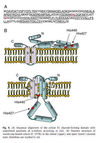
The cytocoxic domain of Colicin R1 forms a channel in the membrane of the E. coli cell once it has passed into the cell. The opening of the channel is controlled in a lipid-dependent manner by a histadine residue at 440. The open channel changes the rate of increase in the membrane conductance, and this change is maximum at an acidic pH. Once bound, there is a pH shift from 4 to 6 on both sides of the membrane. This causes a large increase in the trans-membrane current.
Alkalinization-induced weakening of the electrostatic interactions between colicin and the membrane surface facilitates conformational changes required for the transition of membrane-bound colicin molecules to an active channel state. The channel activity of colicin E1 is not monotonically dependent on the magnitude of the negative surface potential of the target membrane.
Introducing a His440Ala mutation (changing a positive residue to a neutral one) eliminates the pH-shift effect, showing that the change is associated with deprotonation of the His residue, which occurs at an acidic pH. An H427A mutation behaves similarly to the wild type, indicating that it is the deprotonisation of His440 that induces the alkalinization activation. His 440 is located near to the lipid head groups of the bilayer, and His 427 is in the helix that is translocated to the trans-side of the membrane when the channel is open.
The in vitro activity of channel-forming colicins is largest at an acidic ambient pH - colicin E1 is maximum at less than pH5, with a membrane potential of -60mV. This has been ascribed to the inecreased binding of colicin moveles bearing a greater net positive charge to the negatively charged phospholipid surface of the membrane and protein unfolding which involves a massive conformational change.
When the membrane channel lowers, there is a decrease in channel activity, because of electrostatic interactions strong enough to limit the conformational freedom required to insert the colicin from its surface bound state into the E. coli membrane. The activity is also inhibited by calcium ions. This is only observed with negatively charged phospholipid membranes.
References
- ↑ Masi M, Vuong P, Humbard M, Malone K, Misra R. Initial steps of colicin E1 import across the outer membrane of Escherichia coli. J Bacteriol. 2007 Apr;189(7):2667-76. Epub 2007 Feb 2. PMID:17277071 doi:10.1128/JB.01448-06
- ↑ Sobko AA, Rokitskaya TI, Kotova EA. Histidine 440 controls the opening of colicin E1 channels in a lipid-dependent manner. Biochim Biophys Acta. 2009 Sep;1788(9):1962-6. Epub 2009 Jun 26. PMID:19560438 doi:10.1016/j.bbamem.2009.06.017

