We apologize for Proteopedia being slow to respond. For the past two years, a new implementation of Proteopedia has been being built. Soon, it will replace this 18-year old system. All existing content will be moved to the new system at a date that will be announced here.
Journal:PLoS ONE:1
From Proteopedia
(Difference between revisions)

| Line 1: | Line 1: | ||
| + | [[Image:Polio.png|left|200px]] | ||
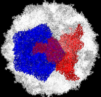
| + | Complete Poliovirus 2 Viron based on PDB entry [[1eah]], <font color='red'><b>example of 3-fold symmetry is in red</b></font>, <font color='blue'><b>example of 5-fold symmetry is in blue</b></font>. | ||
<StructureSection load='Jmol1.pdb' size='500' side='right' scene='Journal:PLoS_ONE:1/F1/1' caption=''> | <StructureSection load='Jmol1.pdb' size='500' side='right' scene='Journal:PLoS_ONE:1/F1/1' caption=''> | ||
=== ANTIVIRAL ACTIVITY of 3(2H)- and 6-CHLORO-3(2H)-ISOFLAVENES AGAINST HIGHLY DIVERGED, NEUROVIRULENT VACCINE-DERIVED, TYPE 2 POLIOVIRUS SEWAGE ISOLATES === | === ANTIVIRAL ACTIVITY of 3(2H)- and 6-CHLORO-3(2H)-ISOFLAVENES AGAINST HIGHLY DIVERGED, NEUROVIRULENT VACCINE-DERIVED, TYPE 2 POLIOVIRUS SEWAGE ISOLATES === | ||
| Line 4: | Line 6: | ||
<big>Lester M. Shulman, Danit Sofer, Yossi Manor, Ella Mendelson, Jean Balanant, Anna Laura Salvati, Francis Delpeyroux, Lucia Fiore</big><ref>DOI</ref> | <big>Lester M. Shulman, Danit Sofer, Yossi Manor, Ella Mendelson, Jean Balanant, Anna Laura Salvati, Francis Delpeyroux, Lucia Fiore</big><ref>DOI</ref> | ||
<hr/> | <hr/> | ||
| - | <b>Molecular Tour</b><br> | + | <b>Molecular Tour</b><br> |
| - | + | ||
| - | + | ||
Poliovirus is a member of the ''Picornaviridae''. Like other members of the ''Picornaviridae'', poliovirus RNA is encapsulated in an icosahedral structure with axes of <scene name='Journal:PLoS_ONE:1/F43/2'>three-fold</scene> and <scene name='Journal:PLoS_ONE:1/F45/1'>five-fold</scene> symmetry formed from 60 <scene name='Journal:PLoS_ONE:1/F1/3'>capsomeres containing one copy</scene> each of viral capsid proteins VP1 (light blue), VP2 (pale green), VP3 (light orange) and VP4(light magenta). The binding site for the human poliovirus receptor is located <scene name='Journal:PLoS_ONE:1/F45/2'>in a canyon at the five-fold axis of symmetry</scene>. The VP1 of picornaviruses contain a hydrophobic pocket that is accessed through this canyon. This pocket is normally occupied by <scene name='Journal:PLoS_ONE:1/F45/3'>pocket factors, sphingosine-like molecules including palmitic and myristic acids and hydrophobic compounds, that stabilize the capsid and whose removal is a necessary prerequisite for uncoating</scene>. The <scene name='Journal:PLoS_ONE:1/F45/4'>broad-spectrum antiviral agent SCH48973</scene> is observed binding in a pocket within the beta-barrel of VP1, in approximately the same location that natural pocket factors bind to polioviruses. SCH48973 forms predominantly hydrophobic interactions with the pocket residues. | Poliovirus is a member of the ''Picornaviridae''. Like other members of the ''Picornaviridae'', poliovirus RNA is encapsulated in an icosahedral structure with axes of <scene name='Journal:PLoS_ONE:1/F43/2'>three-fold</scene> and <scene name='Journal:PLoS_ONE:1/F45/1'>five-fold</scene> symmetry formed from 60 <scene name='Journal:PLoS_ONE:1/F1/3'>capsomeres containing one copy</scene> each of viral capsid proteins VP1 (light blue), VP2 (pale green), VP3 (light orange) and VP4(light magenta). The binding site for the human poliovirus receptor is located <scene name='Journal:PLoS_ONE:1/F45/2'>in a canyon at the five-fold axis of symmetry</scene>. The VP1 of picornaviruses contain a hydrophobic pocket that is accessed through this canyon. This pocket is normally occupied by <scene name='Journal:PLoS_ONE:1/F45/3'>pocket factors, sphingosine-like molecules including palmitic and myristic acids and hydrophobic compounds, that stabilize the capsid and whose removal is a necessary prerequisite for uncoating</scene>. The <scene name='Journal:PLoS_ONE:1/F45/4'>broad-spectrum antiviral agent SCH48973</scene> is observed binding in a pocket within the beta-barrel of VP1, in approximately the same location that natural pocket factors bind to polioviruses. SCH48973 forms predominantly hydrophobic interactions with the pocket residues. | ||
Revision as of 15:29, 27 March 2011
Complete Poliovirus 2 Viron based on PDB entry 1eah, example of 3-fold symmetry is in red, example of 5-fold symmetry is in blue.
| |||||||||||
- ↑ DOI
This page complements a publication in scientific journals and is one of the Proteopedia's Interactive 3D Complement pages. For aditional details please see I3DC.

