User:Dan Huettner/Sandbox 1
From Proteopedia
| Line 22: | Line 22: | ||
===STRUCTURE AND ACTIVE SITE OF HIV-1 PROTEASE=== | ===STRUCTURE AND ACTIVE SITE OF HIV-1 PROTEASE=== | ||
| + | |||
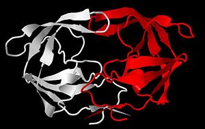
| + | The first structures of HIV-1 protease were reported in 1989 revealing its homodimeric structure consisting of two identical monomers, each made up of 99 amino acids residues, related by a two-fold axis of symmetry (Spinelli et al., 1991). The secondary structure of each monomer contains antiparallel beta-sheets and a single alpha-helix (Figure 3). | ||
| + | |||
| + | Flexible flaps form from an extended turn of a beta sheet (beta hairpin loop) that covers the active site (Figure 3) (Louis et al., 2007). Incidentally, the active site tunnel is normally too narrow for the polyprotein to fit, as depicted in Figure 4. However, the solution is the two flexible protein flaps (amino acid resides 45-55 on each monomer) that can move to allow the peptide to enter the tunnel active site (Toth and Borics, 2006). These flaps undergo a dramatic conformational change from open to closed states to bind the substrate in the proper conformation for catalytic cleavage. The highly flexible tips of the flaps are glycine rich, which curl inside the cleft as the tunnel expands, burying many hydrophobic residues and exposing electronegative active site, while widening the tunnel enough for the substrate to enter (Toth and Borics, 2006). | ||
| + | |||
<Structure load='1HSG' size='500' frame='true' align='right' caption='HIV-1 Protease' scene='Insert optional scene name here' /> | <Structure load='1HSG' size='500' frame='true' align='right' caption='HIV-1 Protease' scene='Insert optional scene name here' /> | ||
| + | |||
===DRUG TARGETS=== | ===DRUG TARGETS=== | ||
Revision as of 04:54, 29 April 2011
Contents |
HIV-1 PROTEASE
INTRODUCTION

Acquired immune deficiency syndrome (AIDS) is arguably of the most prominent and deleterious pandemics of the modern era. AIDS is a condition in humans that causes progressive failure of the immune system, leaving individuals susceptible to opportunistic infections and cancers. The failure of the immune system results from viral assault on the human body’s white blood cells. Since the middle of the 20th century, the disease has spread rapidly in a near ubiquitous fashion, and now the world is on the brink of the fourth decade of the AIDS epidemic.
In the United States, the Center for Disease Control (CDC) first recognized AIDS during the summer of 1981 (Gallo, 2002). Subsequently in 1983, the discovery of the sinister “AIDS virus” coined it as the human immunodeficiency virus (HIV). According to the 2010 UNAIDS Global Report, in 2009, there were 33.3 million people (adults and children) living with HIV, and 1.8 million AIDS related deaths that year, which leads to a deplorable grand total of roughly 30 million deaths due to AIDS since the HIV virus began ravaging the world.
VIROLOGY
The HIV is classified in the Retroviridae family, and more specifically belongs to the genus Lentivirus – slow viruses characterized by a long incubation period, which typically result in long-duration illnesses (Lévy, 1993). Retroviruses (retro is the Latin prefix for “backward”) uniquely possess an RNA genome along with an RNA-dependent DNA polymerase, reverse transcriptase, which is an enzyme that can remarkably direct the synthesis of DNA from RNA (Nelson and Cox, 2008).
The HIV retrovirus has a fairly simple life cycle. The infection begins with the fusion of the viral and host membranes of essential immune cells, typically T-cells and macrophages. Reverse transcriptase then produces DNA from the virus RNA genome, which is then inserted into the host's chromosome by another key enzyme, integrase. At this point the viral genome can become dormant indefinitely. Alternatively, it undergoes transcription and translation into proteins and RNA, required for the new virus. Much of the viral genes are translated initially into precursor polyproteins, which are ultimately cleaved by a final key enzyme of the virus, HIV protease (HIV-1 protease is specific to the HIV-1 class) (Figure 2). The cleavage of the polyproteins into individual proteins may occur during assembly and/or after budding of the immature virus from the host cell (Gelderblom, 1997). These final steps completed by HIV protease are absolutely critical to the maturation of the new virus.
Since the HIV retrovirus contains enzymes imperative for its life cycle that are unique from humans, these are potential inhibitory drug targets to treat infection and to prevent the progression to AIDS. The usual, most effective treatment for HIV infection is a cocktail of drugs that inhibit fusion of the virus and the key enzymes reverse transcriptase, integrase, and protease.
STRUCTURE AND ACTIVE SITE OF HIV-1 PROTEASE
The first structures of HIV-1 protease were reported in 1989 revealing its homodimeric structure consisting of two identical monomers, each made up of 99 amino acids residues, related by a two-fold axis of symmetry (Spinelli et al., 1991). The secondary structure of each monomer contains antiparallel beta-sheets and a single alpha-helix (Figure 3).
Flexible flaps form from an extended turn of a beta sheet (beta hairpin loop) that covers the active site (Figure 3) (Louis et al., 2007). Incidentally, the active site tunnel is normally too narrow for the polyprotein to fit, as depicted in Figure 4. However, the solution is the two flexible protein flaps (amino acid resides 45-55 on each monomer) that can move to allow the peptide to enter the tunnel active site (Toth and Borics, 2006). These flaps undergo a dramatic conformational change from open to closed states to bind the substrate in the proper conformation for catalytic cleavage. The highly flexible tips of the flaps are glycine rich, which curl inside the cleft as the tunnel expands, burying many hydrophobic residues and exposing electronegative active site, while widening the tunnel enough for the substrate to enter (Toth and Borics, 2006).
|
