This old version of Proteopedia is provided for student assignments while the new version is undergoing repairs. Content and edits done in this old version of Proteopedia after March 1, 2026 will eventually be lost when it is retired in about June of 2026.
Apply for new accounts at the new Proteopedia. Your logins will work in both the old and new versions.
Group:MUZIC:Plectin
From Proteopedia

| Line 1: | Line 1: | ||
| - | |||
<Structure load='1sh6' size='500' frame='true' align='right' caption='Insert caption here' scene='Insert optional scene name here' /> | <Structure load='1sh6' size='500' frame='true' align='right' caption='Insert caption here' scene='Insert optional scene name here' /> | ||
Revision as of 07:43, 24 June 2011
|
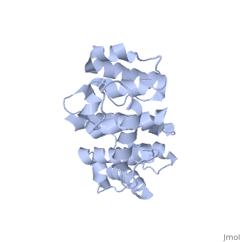
Overall structure
Plectin can be divided in three main sections; a central coiled-coil rod domain, N and C-terminal globular region and exhibits a dumbbell like structure. C-terminal region is composed of 6 homologous repeating domains, and this region has a role in binding to intermediate filaments such as vimentin and cytokeratin [1]. N-terminal globular region contains actin binding domain (ABD) comprising two calponin homology (CH) domains and N-terminal arm, which varies among isoforms[2].
- ↑ Foisner R, Wiche G. Structure and hydrodynamic properties of plectin molecules. J Mol Biol. 1987 Dec 5;198(3):515-31. PMID:3430617
- ↑ Fuchs P, Zorer M, Rezniczek GA, Spazierer D, Oehler S, Castanon MJ, Hauptmann R, Wiche G. Unusual 5' transcript complexity of plectin isoforms: novel tissue-specific exons modulate actin binding activity. Hum Mol Genet. 1999 Dec;8(13):2461-72. PMID:10556294
Proteopedia Page Contributors and Editors (what is this?)
Jae-Geun Song, Alexander Berchansky, Nikos Pinotsis, Jaime Prilusky, Michal Harel
