This old version of Proteopedia is provided for student assignments while the new version is undergoing repairs. Content and edits done in this old version of Proteopedia after March 1, 2026 will eventually be lost when it is retired in about June of 2026.
Apply for new accounts at the new Proteopedia. Your logins will work in both the old and new versions.
Group:MUZIC:Telethonin
From Proteopedia

| Line 1: | Line 1: | ||
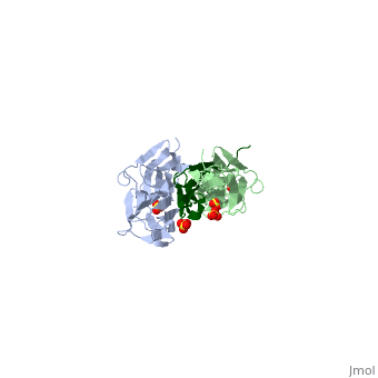
| - | <Structure load='1ya5' size='500' frame='true' align='right' caption='Zou et al. (2006)' scene='<scene name='User:Marcia_Ivonne_Pena_Paz/workbench/Telethonin/Close_tcap/1' | + | <Structure load='1ya5' size='500' frame='true' align='right' caption='Zou et al. (2006)' scene='<scene name='User:Marcia_Ivonne_Pena_Paz/workbench/Telethonin/Close_tcap/1'>' /> |
== Telethonin == | == Telethonin == | ||
Revision as of 07:50, 4 July 2011
' />
TelethoninAlso known as T-Cap or Titin Cap protein. It is a small protein of 19kDa, 167 amino acids. Predominantly expressed in striated muscle. It is a structural protein of the muscle; it is associated to the Z-disc in the sarcomere. It acts as link between titin and other proteins implicated in muscle structure and signalling. Gene regulation and expressionIt is encoded by Tcap gene, in mice (Mus musculus) and humans (Homo sapiens). In mice it is located in chromosome 11, in humans in the long arm of chromosome 17. Tcap is encoded by two exons, and has non-conserved intragenic sequences. The gene is flanked by Stard3 upstream separated by 2,8kb, and Pnmt1 downstream separated by 1,7kb. It has three conserved E-box elements at -103bp (E1), -272bp (E2), and -2067bp (E3). For the full activation of the gene the regulation of E1 is highly important. MyoD plays an important role in this regulation, and from myogenin in late differentiation of myoblasts. [1]
At the transcriptional level it is thought that there is only one isoform ot Tcap, and it is one of the most abundant transcripts in skeletal muscle [2]. It does not have different levels of expression in different type of skeletal muscle; levels of expression of Tcap are lower in neonatal compared to adult striated muscle. The transcript is accumulated in a linear pattern similar to that of the myosin heavy chain [3]. In these same studies it was reported that dennervation lead to decrease in expression of Tcap, suggesting that locomotor activity is also a regulator for its maintenance. Tcap proteinTelethonin protein is found mostly in skeletal and cardiac muscle. It is one of the major components of the sarcomere, it is localized to the Z-disc. Studies on telethonin structure by Zou et al. [4] report that it is formed of five stranded antiparallel β-sheet extended by two wing-shaped β-hairpin motifs (A-B, C-D). And these two motifs are related by an approximate two-fold symmetry, which generates an almost perfect palindromic arrangement. The structure of telethonin was determined using X-ray crystallography [5],[6] . The shape and architecture of the complex of titin/telethonin was studied by small-angle- X-ray scattering (SAXS) and then compared to the crystallographic models. They also used in-vitro experiments to follow the formation of the complex in non-myogenic Cos1 cells, in order to understand if the assemblage is possible [7] This symmetry of telethonin permits its interaction with titin. Both are assembled in an antiparallel sandwich in a (2:1, titin:telethonin). Titin N-terminal domains Z1 and Z2 interact with the N-terminal region of telethonin (residues 1-53). Telethonin mediates in the antiparallel assembly of the two Z1Z2domains. In early differenciating myocyets titin C-terminal and telethonin co-localize and titin kinase is close to telethonin C-terminal, and it is phosphorilated. This phosphorilation is involved in the reorganization of the cytoskeleton during myofibrillogenesis. [8] This co-localization is not seen in adult myofibrils, titin kinase is reported to localize in the M-band [9]; It was also informed that telethonin interacts with other proteins including: Potassium channel subuint miK/isk [10], ankyrin1, and Z-disc proteins FATZ, Calsarcin-3 [11], [12], Ankrd2,[13] and MLP. [14] MLP-Telethonin complex is part of the mechanical stretch sensor machinery in cardiac muscle. The binding site of telethonin and MLP is adjacent to the titing binding domain. This interaction is critical for maintaining normal cardiac function. MLP is required for the stabilization of telethonin-titin complex, this mechanism is not yet clear. The interaction between Ankrd2 and telethonin is been proposed as a sensor of muscle stress/stretch and transmission of the signal to the nucleus regulating gene expression. [15] Telethonin is also involved in signalling processes that regulate muscle development. A feed back loop is formed MRFs (MyoD, myogenin, Myf5) regulating Tcap gene expression; telethonin interacts with myostatin inhibiting it. So it regulates MyoD through Myostatin – Smad3 pathway. [16]. Image:Telethonin Myostatin.jpg Pathologies associated to telethoninDifferent mutations in telethonin have been associated to several myopathies. Mutations that lead to limb-girdle muscular dystrophy type 2G (LGMD2G) [17], to hypertrophic cardiopathy., [18], dilated cardiomyopathy. A frameshift mutation in Tcap gene leads to a deletion of telethonin C-terminal region, loosing the region for phosphorilation control [19], as it was observed in few brazilian families. Defects in the MLP-Tcap association are linked to human dilated cardiomyopathy and heart failure (Knöll 2002). Mutations that affect ability of MLP to interact with telethonin result in the loss of telethonin binding, facilitating its misslocalization from the complex with titin, this brings to defects in the Z-disc and progression of dilated cardiomyopathy. Knöll et al. conclude that genetic mutations causing a defectuos interaction of telethonin with MLP can lead to a development of human dilated cardiomyopathy through modifications in the conformation and function of titin. It was reported that in 10 cases of neurogenic atrophy there was a decrease staining for telethonin in type II fibers, in early stages of fiber atrophy, [20] indicating a selective downregulation of telethonin. These observations can be related to in vivo studies in rats in which after short term dennervation (two days), Tcap transcript is reduced about 50% in skeletal muscle. [21].
References
| ||||||||||||

