User:Gourinchas Geoffrey/Sandbox 205
From Proteopedia
| Line 2: | Line 2: | ||
[[Image:Erythropoietin.png|200px|left|thumb|Erythropoeitin]] <Structure load='1buy' size='500' frame='true' align='right' caption='Molecule of Erythropoietin' scene='User:Gourinchas_Geoffrey/Sandbox_205/Picture_of_presentation_n3_bla/1' /> | [[Image:Erythropoietin.png|200px|left|thumb|Erythropoeitin]] <Structure load='1buy' size='500' frame='true' align='right' caption='Molecule of Erythropoietin' scene='User:Gourinchas_Geoffrey/Sandbox_205/Picture_of_presentation_n3_bla/1' /> | ||
| - | The [http://en.wikipedia.org/wiki/Erythropoietin Erythropoietin] hormone is a glycoprotein which is involved in [http://en.wikipedia.org/wiki/Erythropoiesis Erythropoiesis], which is the red blood cells production. | + | The [http://en.wikipedia.org/wiki/Erythropoietin Erythropoietin] hormone is a glycoprotein which is involved in [http://en.wikipedia.org/wiki/Erythropoiesis Erythropoiesis], which is the red blood cells production <ref>ANNALS Of ANATOMY, Erythropoietin in the control of red cell production, Wolfgang Jelkmann and Eric Metzen, Institut fiir Physiologie. Medizinische Uni versitat zu Liibeck, Ratzeburger Allee 160, |
| + | D-23538 Liibeck</ref>. | ||
It allows the differenciation of the erythrocyte precursors in the bone marrow. Thus, Epo has a fundamental biological role in the regulation of the production of red blood cells and, thus, at the level of the oxygenation of the blood. | It allows the differenciation of the erythrocyte precursors in the bone marrow. Thus, Epo has a fundamental biological role in the regulation of the production of red blood cells and, thus, at the level of the oxygenation of the blood. | ||
| Line 50: | Line 51: | ||
The gene of this hormone code for a protein of 193 amino acids. The signal peptide which is constituted to 27 amino acids is split and moreover the hormone loses its terminal Arginine in position 166. Thus active Erythropoietin is constituted to 165 amino acids. | The gene of this hormone code for a protein of 193 amino acids. The signal peptide which is constituted to 27 amino acids is split and moreover the hormone loses its terminal Arginine in position 166. Thus active Erythropoietin is constituted to 165 amino acids. | ||
The molecule of Erythropoietin is strongly glycosylated. It has 3 sites of N-Glycosylation and one of O-Glycosylation. When Erythropoietin is completely glycosylated this hormone weight 30.4kD, among which 40 % are represented by carbohydrates. It also possesses 14 sialic acids. | The molecule of Erythropoietin is strongly glycosylated. It has 3 sites of N-Glycosylation and one of O-Glycosylation. When Erythropoietin is completely glycosylated this hormone weight 30.4kD, among which 40 % are represented by carbohydrates. It also possesses 14 sialic acids. | ||
| - | The in vivo biological action is dependent on the glycosylation: indeed if the molecule loses its acids sialiques, stripped galactoses are recognized by the specific hepatic receptor, also the molecule is evacuated then quickly by the circulation. On the contrary,if the Erythropoietin is completely no glycosylated, it is going to aggregated and to become completely inactive in vivo. | + | The in vivo biological action is dependent on the glycosylation <ref>http://www.sciencedirect.com/science/article/pii/S0022283611008394</ref>: indeed if the molecule loses its acids sialiques, stripped galactoses are recognized by the specific hepatic receptor, also the molecule is evacuated then quickly by the circulation. On the contrary,if the Erythropoietin is completely no glycosylated, it is going to aggregated and to become completely inactive in vivo. |
The tertiary structure of this hormone is composed to 4 alpha-helix: αA, αB, αC and αD which are associated the some in front of the others. | The tertiary structure of this hormone is composed to 4 alpha-helix: αA, αB, αC and αD which are associated the some in front of the others. | ||
| Line 63: | Line 64: | ||
In the second interaction site, the Methionine 150 allows van der waals interactions with Arginine 10, the Valine 11 and the Arginine 14 of Erythropoietin. | In the second interaction site, the Methionine 150 allows van der waals interactions with Arginine 10, the Valine 11 and the Arginine 14 of Erythropoietin. | ||
| - | The hydrophobic interaction include 11 hydrogen bonds between αA and αC of Erythropoietin and his receptor. | + | The hydrophobic interaction include 11 hydrogen bonds between αA and αC of Erythropoietin and his receptor. <ref>4<ref/> |
| Line 94: | Line 95: | ||
<references/> http://biology.kenyon.edu/BMB/Chime2/2004/eryth/FRAMES/start.htm | <references/> http://biology.kenyon.edu/BMB/Chime2/2004/eryth/FRAMES/start.htm | ||
| + | |||
| + | <references/>J. Mol. Biol. (2011) 412, 536–550 | ||
Revision as of 15:36, 23 December 2011
Contents |
HUMAN ERYTHROPOIETIN

|
The Erythropoietin hormone is a glycoprotein which is involved in Erythropoiesis, which is the red blood cells production [1]. It allows the differenciation of the erythrocyte precursors in the bone marrow. Thus, Epo has a fundamental biological role in the regulation of the production of red blood cells and, thus, at the level of the oxygenation of the blood.
Biological role
The gene of the erythropoietin was cloned at the man's, the mouse and the monkey. It is located at the man's on the chromosome 7 in the region q21. It contains 5 exons and 4 introns. The gene is very preserved according to the species. It exists 94 % of homology between the human gene and the monkey gene and 80 % of homology enters the murin gene and the human gene. L'Erythropoietin is a glycosylated hormone mainly secreted in the kidneys (90%) at the level of the cortex and of external medullary, and little in the liver. This hormone acts on the target cells by the intermediary of a specific receptor essentially on the erythroblastic progenitor, and a little on megacaryocyte progenitor. Through experiences of connection with the market hormone, it was demonstrated the existence of a single receptor (high affinity) or two types of receptor of high and low affinity. After the binding with its receptor, the Erythropoietin specifically leads the phosphorylation of the receptor on the tyrosine, from the first minutes which follow its connection.
The secretion of this hormone depend of the partial pressure of dioxygen in the kidney cells. The rate of Erythropoietin increases when there is a tissular hypoxie which origin can be anemic, secondary in an obstructive lung syndrome, in a left-right cardiac shunt or, much more rarely, in an abnormality of the affinity of the haemoglobin for oxygen. Thus needs of dioxygen of the tissues increase and thus the secretion of Erythropoietin is stimulated.
On the contrary, an increase of the pressure of dioxygen decrease the secretion of Erythropoietin.
To increase the number of red blood cells, this hormone stimulate the proliferation of stem cells precursor of erythrocyte in the bone marrow which will became red blood cells.
For regulate the expression of the Erythropoietin, there are activating regulating sequences in 3 ' of the gene as well as the sequences in 5 ' which are specific of tissue. On the other hand, recent works suggest that a heminique protein would play a regulating role on the transcription: if the tissular oxygenation is adequate, this protein fixes the oxygen and inhibits the transcription of Erythropoietin.
In the absence of oxygen, the deoxygenated shape of this heminique protein could lead the production of the Erythropoietin. The glycosylation of this hormone allows his solubilisation in the blood and his carry towards his target, the bone marrow.
Structure
| |||||||||||
αα
Application in medicine
See also
crystal structure of human erythropoietin complexed to its receptor at 1.9 angstroms.
complex between the extracellular domain of erythropoietin (epo) receptor [ebp and an inactive peptide [emp33] contains 3,5-dibromotyrosine in position 4 (denoted dby)]
complex between the extracellular domain of erythropoietin (epo) receptor [ebp and an agonist peptide [emp1]]
Native structure of the extracellular domain of erythropoietin (epo) receptor [ebp]
References
- ↑ ANNALS Of ANATOMY, Erythropoietin in the control of red cell production, Wolfgang Jelkmann and Eric Metzen, Institut fiir Physiologie. Medizinische Uni versitat zu Liibeck, Ratzeburger Allee 160, D-23538 Liibeck
- ↑ http://www.sciencedirect.com/science/article/pii/S0022283611008394
- ↑ 4<ref></ref>
Optimal angle for Erythropoietin binding to his receptor.
Syed and al. in 1998 have showed that Erythropoietin optimal binds at his receptor if only there is an angle of 120° between the two sites of the receptor <ref>http://biology.kenyon.edu/BMB/Chime2/2004/eryth/FRAMES/start.htm</li></ol></ref>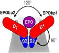 Erythropoeitin with his receptor

J. Mol. Biol. (2011) 412, 536–550
