This old version of Proteopedia is provided for student assignments while the new version is undergoing repairs. Content and edits done in this old version of Proteopedia after March 1, 2026 will eventually be lost when it is retired in about June of 2026.
Apply for new accounts at the new Proteopedia. Your logins will work in both the old and new versions.
Sandbox SouthUniversity1
From Proteopedia
| Line 19: | Line 19: | ||
The reason that this flavone is bound so tightly to the enzyme is that it's shape and electronic charge are complementary to the binding pocket. This is examined <scene name='Sandbox_SouthUniversity1/2hi4_blue_transparent_ribbons/1'>next.</scene> | The reason that this flavone is bound so tightly to the enzyme is that it's shape and electronic charge are complementary to the binding pocket. This is examined <scene name='Sandbox_SouthUniversity1/2hi4_blue_transparent_ribbons/1'>next.</scene> | ||
| - | First examine the shape of the VDW area <scene name='Sandbox_SouthUniversity1/2hi4_bl_transp_rib_flav_vdw/1'>around the flavone.</scene> | + | First examine the shape of the VDW area <scene name='Sandbox_SouthUniversity1/2hi4_bl_transp_rib_flav_vdw/1'>around the flavone.</scene> The reddish areas show places where there are particularly close contacts with the binding pocket. These contacts can be caused by ionic, hydrophobic, or hydrogen bonding. Now we will examine what exactly is responsible for the tight binding. |
| + | |||
| + | Lets remove the rest of the protein, so we can better see these interactions. | ||
First examine the shape of the <scene name='Sandbox_SouthUniversity1/2hi4_bl_transp_ribbon_flav_spc/1'>flavone </scene>bound to the protein. | First examine the shape of the <scene name='Sandbox_SouthUniversity1/2hi4_bl_transp_ribbon_flav_spc/1'>flavone </scene>bound to the protein. | ||
Current revision
==Selectivity of Drug Metabolism by Cytochrome P450 Enzymes==
| |||||||||||
Draft: <illustration of residues in active site> Draft animation Draft animation
draft: <insert quiz here on electrostatic and steric factors>
draft: <insert animations here contrasting the shape of the active site of CYP3A4 and e.g. CYP2E1>
draft: <insert examples of substrates for 2 different CYPs, overlay them on top of each other illustrate the molecular commonalities>
draft <illustrate the size of the binding pockets and their electronic character>
draft test of scene names:
<Draft Quiz>
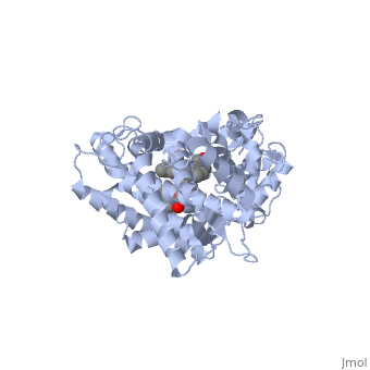
References The structure of CYP1A2 shown above is found in the Research Collaboration of Structural Bioinformatics Protein Database as entry 2HI4.
