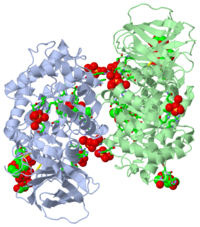
Acid beta-glucosidase with N-butyl-deoxynojirimycin
From Proteopedia
(Difference between revisions)
(New page: <StructureSection load="2fcj" size="400" color="" frame="true" spin="on" Scene='2v3d/Cv/1' align="right" caption= > 200px <!-- The line below this paragraph, co...) |
|||
| Line 2: | Line 2: | ||
[[Image:2v3d.png|left|200px]] | [[Image:2v3d.png|left|200px]] | ||
| - | |||
| - | <!-- | ||
| - | The line below this paragraph, containing "STRUCTURE_2v3d", creates the "Structure Box" on the page. | ||
| - | You may change the PDB parameter (which sets the PDB file loaded into the applet) | ||
| - | or the SCENE parameter (which sets the initial scene displayed when the page is loaded), | ||
| - | or leave the SCENE parameter empty for the default display. | ||
| - | --> | ||
| - | |||
===ACID-BETA-GLUCOSIDASE WITH N-BUTYL-DEOXYNOJIRIMYCIN ([[2v3d]])=== | ===ACID-BETA-GLUCOSIDASE WITH N-BUTYL-DEOXYNOJIRIMYCIN ([[2v3d]])=== | ||
(see also [[Treatment of Gaucher disease]]) | (see also [[Treatment of Gaucher disease]]) | ||
| - | <!-- | ||
| - | The line below this paragraph, {{ABSTRACT_PUBMED_17666401}}, adds the Publication Abstract to the page | ||
| - | (as it appears on PubMed at http://www.pubmed.gov), where 17666401 is the PubMed ID number. | ||
| - | --> | ||
{{ABSTRACT_PUBMED_17666401}} | {{ABSTRACT_PUBMED_17666401}} | ||
{{Clear}} | {{Clear}} | ||
| - | <scene name='2v3d/Al/3'>Superimposition</scene> of the <font color='red'><b>native human acid β-glucosidase, expressed in cultured plant cells (pGlcCerase,</b></font> [[2v3f]]) on those of <font color='darkmagenta'><b>N-butyl-deoxynojirimycin/pGlcCerase</b></font> ( | + | <scene name='2v3d/Al/3'>Superimposition</scene> of the <font color='red'><b>native human acid β-glucosidase, expressed in cultured plant cells (pGlcCerase,</b></font> [[2v3f]]) on those of <font color='darkmagenta'><b>N-butyl-deoxynojirimycin/pGlcCerase</b></font> ([[2v3d]]), <font color='lime'><b>N-nonyl-deoxynojirimycin/pGlcCerase</b></font> ([[2v3e]]), and <font color='cyan'><b>isofagomine/deglycosylated Cerezyme</b></font> (IFG/DG-Cerezyme, [[2nsx]]) reveals significant structural identity, neither of these ligands causes structural changes upon binding to the enzyme. The imino sugar of <font color='magenta'><b>N-butyl-deoxynojirimycin</b></font> <scene name='2v3d/Al/10'>(NB-DNJ)</scene> forms 7 hydrogen bonds and also makes several hydrophobic interactions with side chains of <font color='darkmagenta'><b>active site residues</b></font> ('''2v3d'''). The crystal structure of <font color='lime'><b>pGlcCerase in complex</b></font> with <font color='orange'><b>N-nonyl-deoxynojirimycin</b></font> <scene name='2v3d/Al/11'>(NN-DNJ)</scene> ([[2v3e]]) is very similar to that of <font color='magenta'><b>NB-DNJ</b></font>/<font color='darkmagenta'><b>pGlcCerase</b></font>. The exception is that longer chain of <font color='orange'><b>NN-DNJ</b></font> interacts with 2 additional residues Leu241 (<font color='lime'><b>labeled lime</b></font>) and Leu314 of symmetrically related monomer (not shown). Comparison of the structures of NB-DNJ/pGlcCerase ([[2v3d]]) and NN-DNJ/pGlcCerase ([[2v3e]]) with that of <scene name='2v3d/Nsx/2'>IFG/DG-Cerezyme</scene> ([[2nsx]]) shows that the pyranose-like ring forms a same number of hydrogen bonds with the enzyme in all three cases ([[2v3d]], [[2v3e]], and [[2nsx]]). |
==About this Structure== | ==About this Structure== | ||
Revision as of 10:47, 11 June 2012
| |||||||||||
Reference
- Brumshtein B, Greenblatt HM, Butters TD, Shaaltiel Y, Aviezer D, Silman I, Futerman AH, Sussman JL. Crystal structures of complexes of N-butyl- and N-nonyl-deoxynojirimycin bound to acid beta-glucosidase: insights into the mechanism of chemical chaperone action in Gaucher disease. J Biol Chem. 2007 Sep 28;282(39):29052-8. Epub 2007 Jul 31. PMID:17666401 doi:10.1074/jbc.M705005200
Categories: Glucosylceramidase | Homo sapiens | Aviezer, D. | Brumshtein, B. | Butters, T D. | Futerman, A H. | Greenblatt, H M. | Shaaltiel, Y. | ISPC, Israel Structural Proteomics Center. | ISPC | Silman, I. | Sussman, J L. | Acid-beta-glucosidase | Alternative splicing | Disease mutation | Gaucher disease | Glycoprotein | Glycosidase | Hydrolase | Lipid metabolism | Lysosome | Membrane | N-butyl-deoxynojirimycin | N-butyl-deoxynojirimycin alternative initiation | Pharmaceutical | Polymorphism | Sphingolipid metabolism

