This old version of Proteopedia is provided for student assignments while the new version is undergoing repairs. Content and edits done in this old version of Proteopedia after March 1, 2026 will eventually be lost when it is retired in about June of 2026.
Apply for new accounts at the new Proteopedia. Your logins will work in both the old and new versions.
2x6v
From Proteopedia
(Difference between revisions)
m (Protected "2x6v" [edit=sysop:move=sysop]) |
|||
| Line 1: | Line 1: | ||
| - | [[Image:2x6v.png|left|200px]] | ||
| - | |||
| - | <!-- | ||
| - | The line below this paragraph, containing "STRUCTURE_2x6v", creates the "Structure Box" on the page. | ||
| - | You may change the PDB parameter (which sets the PDB file loaded into the applet) | ||
| - | or the SCENE parameter (which sets the initial scene displayed when the page is loaded), | ||
| - | or leave the SCENE parameter empty for the default display. | ||
| - | --> | ||
{{STRUCTURE_2x6v| PDB=2x6v | SCENE= }} | {{STRUCTURE_2x6v| PDB=2x6v | SCENE= }} | ||
| - | |||
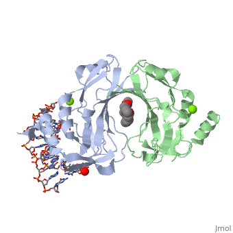
===CRYSTAL STRUCTURE OF HUMAN TBX5 IN THE DNA-BOUND AND DNA-FREE FORM=== | ===CRYSTAL STRUCTURE OF HUMAN TBX5 IN THE DNA-BOUND AND DNA-FREE FORM=== | ||
| - | |||
| - | |||
| - | <!-- | ||
| - | The line below this paragraph, {{ABSTRACT_PUBMED_20450920}}, adds the Publication Abstract to the page | ||
| - | (as it appears on PubMed at http://www.pubmed.gov), where 20450920 is the PubMed ID number. | ||
| - | --> | ||
{{ABSTRACT_PUBMED_20450920}} | {{ABSTRACT_PUBMED_20450920}} | ||
Revision as of 10:09, 11 March 2013
| |||||||
| 2x6v, resolution 2.20Å () | |||||||
|---|---|---|---|---|---|---|---|
| Ligands: | , | ||||||
| Related: | 2x6u | ||||||
| |||||||
| Resources: | FirstGlance, OCA, RCSB, PDBsum | ||||||
| Coordinates: | save as pdb, mmCIF, xml | ||||||
Contents |
CRYSTAL STRUCTURE OF HUMAN TBX5 IN THE DNA-BOUND AND DNA-FREE FORM
Template:ABSTRACT PUBMED 20450920
About this Structure
2x6v is a 4 chain structure with sequence from Homo sapiens. Full crystallographic information is available from OCA.
See Also
Reference
- Stirnimann CU, Ptchelkine D, Grimm C, Muller CW. Structural basis of TBX5-DNA recognition: the T-box domain in its DNA-bound and -unbound form. J Mol Biol. 2010 Jul 2;400(1):71-81. Epub 2010 May 5. PMID:20450920 doi:10.1016/j.jmb.2010.04.052
