We apologize for Proteopedia being slow to respond. For the past two years, a new implementation of Proteopedia has been being built. Soon, it will replace this 18-year old system. All existing content will be moved to the new system at a date that will be announced here.
ExbB
From Proteopedia
(Difference between revisions)
| Line 1: | Line 1: | ||
| + | <StructureSection load='5zfu' size='350' side='right' caption='2-isopropylmalate synthase complex with leucine, glycerol and Zn+2 ion (grey) (PDB code [[5zfu]])' scene=''> | ||
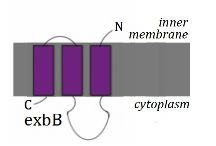
[[Image:ExbB.jpg|300px|right|thumb| The Structure of ExbB<ref name='Kampfenkel'>PMID: 8449962</ref>]] | [[Image:ExbB.jpg|300px|right|thumb| The Structure of ExbB<ref name='Kampfenkel'>PMID: 8449962</ref>]] | ||
| Line 12: | Line 13: | ||
Loss of ExbB function can be partially replaced by TolQ and vice versa - reduced activities of either of these proteins via a mutant form can be reversed by introducing double mutants<ref name='Braun'>PMID: 15205446</ref>. | Loss of ExbB function can be partially replaced by TolQ and vice versa - reduced activities of either of these proteins via a mutant form can be reversed by introducing double mutants<ref name='Braun'>PMID: 15205446</ref>. | ||
| + | </StructureSection> | ||
| + | ==3D structures of ExbB== | ||
| + | |||
| + | Updated on {{REVISIONDAY2}}-{{MONTHNAME|{{REVISIONMONTH}}}}-{{REVISIONYEAR}} | ||
| + | |||
| + | [[5zfu]], [[5zfv]] – ExbD + ExbD peptide – ''Escherichia coli'' – Cryo EM<br /> | ||
== References== | == References== | ||
<references/> | <references/> | ||
Revision as of 07:33, 22 May 2018
| |||||||||||
3D structures of ExbB
Updated on 22-May-2018
5zfu, 5zfv – ExbD + ExbD peptide – Escherichia coli – Cryo EM
References
- ↑ 1.0 1.1 Kampfenkel K, Braun V. Topology of the ExbB protein in the cytoplasmic membrane of Escherichia coli. J Biol Chem. 1993 Mar 15;268(8):6050-7. PMID:8449962
- ↑ 2.0 2.1 Held KG, Postle K. ExbB and ExbD do not function independently in TonB-dependent energy transduction. J Bacteriol. 2002 Sep;184(18):5170-3. PMID:12193634
- ↑ 3.0 3.1 Braun V, Herrmann C. Point mutations in transmembrane helices 2 and 3 of ExbB and TolQ affect their activities in Escherichia coli K-12. J Bacteriol. 2004 Jul;186(13):4402-6. PMID:15205446 doi:10.1128/JB.186.13.4402-4406.2004

