This old version of Proteopedia is provided for student assignments while the new version is undergoing repairs. Content and edits done in this old version of Proteopedia after March 1, 2026 will eventually be lost when it is retired in about June of 2026.
Apply for new accounts at the new Proteopedia. Your logins will work in both the old and new versions.
1xjo
From Proteopedia
(Difference between revisions)
| Line 1: | Line 1: | ||
| - | [[ | + | ==STRUCTURE OF AMINOPEPTIDASE== |
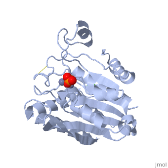
| + | <StructureSection load='1xjo' size='340' side='right' caption='[[1xjo]], [[Resolution|resolution]] 1.75Å' scene=''> | ||
| + | == Structural highlights == | ||
| + | <table><tr><td colspan='2'>[[1xjo]] is a 1 chain structure with sequence from [http://en.wikipedia.org/wiki/Streptomyces_griseus Streptomyces griseus]. Full crystallographic information is available from [http://oca.weizmann.ac.il/oca-bin/ocashort?id=1XJO OCA]. For a <b>guided tour on the structure components</b> use [http://oca.weizmann.ac.il/oca-docs/fgij/fg.htm?mol=1XJO FirstGlance]. <br> | ||
| + | </td></tr><tr><td class="sblockLbl"><b>[[Ligand|Ligands:]]</b></td><td class="sblockDat"><scene name='pdbligand=CA:CALCIUM+ION'>CA</scene>, <scene name='pdbligand=PO4:PHOSPHATE+ION'>PO4</scene>, <scene name='pdbligand=ZN:ZINC+ION'>ZN</scene><br> | ||
| + | <tr><td class="sblockLbl"><b>[[Non-Standard_Residue|NonStd Res:]]</b></td><td class="sblockDat"><scene name='pdbligand=MHO:S-OXYMETHIONINE'>MHO</scene></td></tr> | ||
| + | <tr><td class="sblockLbl"><b>Resources:</b></td><td class="sblockDat"><span class='plainlinks'>[http://oca.weizmann.ac.il/oca-docs/fgij/fg.htm?mol=1xjo FirstGlance], [http://oca.weizmann.ac.il/oca-bin/ocaids?id=1xjo OCA], [http://www.rcsb.org/pdb/explore.do?structureId=1xjo RCSB], [http://www.ebi.ac.uk/pdbsum/1xjo PDBsum]</span></td></tr> | ||
| + | <table> | ||
| + | == Evolutionary Conservation == | ||
| + | [[Image:Consurf_key_small.gif|200px|right]] | ||
| + | Check<jmol> | ||
| + | <jmolCheckbox> | ||
| + | <scriptWhenChecked>select protein; define ~consurf_to_do selected; consurf_initial_scene = true; script "/wiki/ConSurf/xj/1xjo_consurf.spt"</scriptWhenChecked> | ||
| + | <scriptWhenUnchecked>script /wiki/extensions/Proteopedia/spt/initialview01.spt</scriptWhenUnchecked> | ||
| + | <text>to colour the structure by Evolutionary Conservation</text> | ||
| + | </jmolCheckbox> | ||
| + | </jmol>, as determined by [http://consurfdb.tau.ac.il/ ConSurfDB]. You may read the [[Conservation%2C_Evolutionary|explanation]] of the method and the full data available from [http://bental.tau.ac.il/new_ConSurfDB/chain_selection.php?pdb_ID=2ata ConSurf]. | ||
| + | <div style="clear:both"></div> | ||
| + | <div style="background-color:#fffaf0;"> | ||
| + | == Publication Abstract from PubMed == | ||
| + | The X-ray crystal structure of the enzyme Streptomyces griseus aminopeptidase (SGAP) has been determined in its double zinc form to 1.75 A resolution, in its apo-enzyme from (zinc removed) to 2.1 A resolution, and as a mercury replaced derivative to 2.1 A resolution. The structure solution was achieved by single isomorphous replacement with phasing from anomalous scattering (SIRAS), followed by density modification with histogram matching. The protein consists of a central beta-sheet made up of eight parallel and antiparallel strands, surrounded by helices on either side. The active site is located at the carbonyl ends of two middle strands of the beta-sheet region. Two sections of the chain that could not be traced were Glu196 to Arg202, which borders the active site, and the final seven C-terminal residues starting with Gly278. The active site contains two zinc cations, each with similar ligands, at a distance of 3.6 A from each other. An unknown molecule appears to be bound to both zinc ions in the active site at partial occupancy and has been modelled as a phosphate ion. A calcium binding site has also been identified, consistent with the observations that calcium modulates the activity of the enzyme, and increases its heat stability. The mechanism by which the calcium cation modulates enzyme activity is not apparent, since the location of the calcium binding site is approximately 25 A distant from the active site zinc ions. Comparison of the structure of SGAP to other known aminopeptidases shows that the enzyme is most similar to Aeromonas proteolytica aminopeptidase (AAP). Both enzymes share a similar topology, although the overall sequence identity is very low (24% in aligned regions). The coordination of the two active site zinc cations in SGAP resembles that of AAP. These two microbial enzymes differ from bovine lens leucine aminopeptidase (LAP) in both overall structure and in coordination of the two zinc ions. | ||
| - | + | Streptomyces griseus aminopeptidase: X-ray crystallographic structure at 1.75 A resolution.,Greenblatt HM, Almog O, Maras B, Spungin-Bialik A, Barra D, Blumberg S, Shoham G J Mol Biol. 1997 Feb 7;265(5):620-36. PMID:9048953<ref>PMID:9048953</ref> | |
| - | + | From MEDLINE®/PubMed®, a database of the U.S. National Library of Medicine.<br> | |
| - | + | </div> | |
| - | + | ||
| - | + | ||
| - | + | ||
| - | + | ||
==See Also== | ==See Also== | ||
*[[Aminopeptidase|Aminopeptidase]] | *[[Aminopeptidase|Aminopeptidase]] | ||
*[[Streptomyces griseus Aminopeptidase (SGAP)|Streptomyces griseus Aminopeptidase (SGAP)]] | *[[Streptomyces griseus Aminopeptidase (SGAP)|Streptomyces griseus Aminopeptidase (SGAP)]] | ||
| - | + | == References == | |
| - | == | + | <references/> |
| - | < | + | __TOC__ |
| + | </StructureSection> | ||
[[Category: Streptomyces griseus]] | [[Category: Streptomyces griseus]] | ||
[[Category: Barra, D.]] | [[Category: Barra, D.]] | ||
Revision as of 22:34, 28 September 2014
STRUCTURE OF AMINOPEPTIDASE
| |||||||||||