This old version of Proteopedia is provided for student assignments while the new version is undergoing repairs. Content and edits done in this old version of Proteopedia after March 1, 2026 will eventually be lost when it is retired in about June of 2026.
Apply for new accounts at the new Proteopedia. Your logins will work in both the old and new versions.
2h7f
From Proteopedia
(Difference between revisions)
| Line 1: | Line 1: | ||
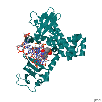
| - | [[ | + | ==Structure of variola topoisomerase covalently bound to DNA== |
| + | <StructureSection load='2h7f' size='340' side='right' caption='[[2h7f]], [[Resolution|resolution]] 2.70Å' scene=''> | ||
| + | == Structural highlights == | ||
| + | <table><tr><td colspan='2'>[[2h7f]] is a 3 chain structure with sequence from [http://en.wikipedia.org/wiki/Variola_virus Variola virus]. Full crystallographic information is available from [http://oca.weizmann.ac.il/oca-bin/ocashort?id=2H7F OCA]. For a <b>guided tour on the structure components</b> use [http://oca.weizmann.ac.il/oca-docs/fgij/fg.htm?mol=2H7F FirstGlance]. <br> | ||
| + | </td></tr><tr><td class="sblockLbl"><b>[[Non-Standard_Residue|NonStd Res:]]</b></td><td class="sblockDat"><scene name='pdbligand=PTR:O-PHOSPHOTYROSINE'>PTR</scene></td></tr> | ||
| + | <tr><td class="sblockLbl"><b>[[Related_structure|Related:]]</b></td><td class="sblockDat">[[2h7g|2h7g]]</td></tr> | ||
| + | <tr><td class="sblockLbl"><b>[[Gene|Gene:]]</b></td><td class="sblockDat">TOP1 ([http://www.ncbi.nlm.nih.gov/Taxonomy/Browser/wwwtax.cgi?mode=Info&srchmode=5&id=10255 Variola virus])</td></tr> | ||
| + | <tr><td class="sblockLbl"><b>Activity:</b></td><td class="sblockDat"><span class='plainlinks'>[http://en.wikipedia.org/wiki/DNA_topoisomerase DNA topoisomerase], with EC number [http://www.brenda-enzymes.info/php/result_flat.php4?ecno=5.99.1.2 5.99.1.2] </span></td></tr> | ||
| + | <tr><td class="sblockLbl"><b>Resources:</b></td><td class="sblockDat"><span class='plainlinks'>[http://oca.weizmann.ac.il/oca-docs/fgij/fg.htm?mol=2h7f FirstGlance], [http://oca.weizmann.ac.il/oca-bin/ocaids?id=2h7f OCA], [http://www.rcsb.org/pdb/explore.do?structureId=2h7f RCSB], [http://www.ebi.ac.uk/pdbsum/2h7f PDBsum]</span></td></tr> | ||
| + | <table> | ||
| + | == Evolutionary Conservation == | ||
| + | [[Image:Consurf_key_small.gif|200px|right]] | ||
| + | Check<jmol> | ||
| + | <jmolCheckbox> | ||
| + | <scriptWhenChecked>select protein; define ~consurf_to_do selected; consurf_initial_scene = true; script "/wiki/ConSurf/h7/2h7f_consurf.spt"</scriptWhenChecked> | ||
| + | <scriptWhenUnchecked>script /wiki/extensions/Proteopedia/spt/initialview01.spt</scriptWhenUnchecked> | ||
| + | <text>to colour the structure by Evolutionary Conservation</text> | ||
| + | </jmolCheckbox> | ||
| + | </jmol>, as determined by [http://consurfdb.tau.ac.il/ ConSurfDB]. You may read the [[Conservation%2C_Evolutionary|explanation]] of the method and the full data available from [http://bental.tau.ac.il/new_ConSurfDB/chain_selection.php?pdb_ID=2ata ConSurf]. | ||
| + | <div style="clear:both"></div> | ||
| + | <div style="background-color:#fffaf0;"> | ||
| + | == Publication Abstract from PubMed == | ||
| + | Although smallpox has been eradicated from the human population, it is presently feared as a possible agent of bioterrorism. The smallpox virus codes for its own topoisomerase enzyme that differs from its cellular counterpart by requiring a specific DNA sequence for activation of catalysis. Here we present crystal structures of the smallpox virus topoisomerase enzyme bound both covalently and noncovalently to a specific DNA sequence. These structures reveal the basis for site-specific DNA recognition, and they explain how catalysis is likely activated by formation of a specific enzyme-DNA interface. Unexpectedly, the poxvirus enzyme uses a major groove binding alpha helix that is not present in the human enzyme to recognize part of the core recognition sequence and activate the enzyme for catalysis. The topoisomerase-DNA complex structures also provide a three-dimensional framework that may facilitate the rational design of therapeutic agents to treat poxvirus infections. | ||
| - | + | Structural basis for specificity in the poxvirus topoisomerase.,Perry K, Hwang Y, Bushman FD, Van Duyne GD Mol Cell. 2006 Aug 4;23(3):343-54. PMID:16885024<ref>PMID:16885024</ref> | |
| - | + | From MEDLINE®/PubMed®, a database of the U.S. National Library of Medicine.<br> | |
| - | + | </div> | |
| - | + | ||
| - | + | ||
| - | + | ||
| - | + | ||
==See Also== | ==See Also== | ||
*[[Topoisomerase|Topoisomerase]] | *[[Topoisomerase|Topoisomerase]] | ||
| - | + | == References == | |
| - | == | + | <references/> |
| - | < | + | __TOC__ |
| + | </StructureSection> | ||
[[Category: DNA topoisomerase]] | [[Category: DNA topoisomerase]] | ||
[[Category: Variola virus]] | [[Category: Variola virus]] | ||
Revision as of 03:58, 29 September 2014
Structure of variola topoisomerase covalently bound to DNA
| |||||||||||