We apologize for Proteopedia being slow to respond. For the past two years, a new implementation of Proteopedia has been being built. Soon, it will replace this 18-year old system. All existing content will be moved to the new system at a date that will be announced here.
1ema
From Proteopedia
(Difference between revisions)
| Line 1: | Line 1: | ||
| - | [[Image:1ema.png|left|200px]] | ||
| - | |||
{{STRUCTURE_1ema| PDB=1ema | SCENE= }} | {{STRUCTURE_1ema| PDB=1ema | SCENE= }} | ||
| - | |||
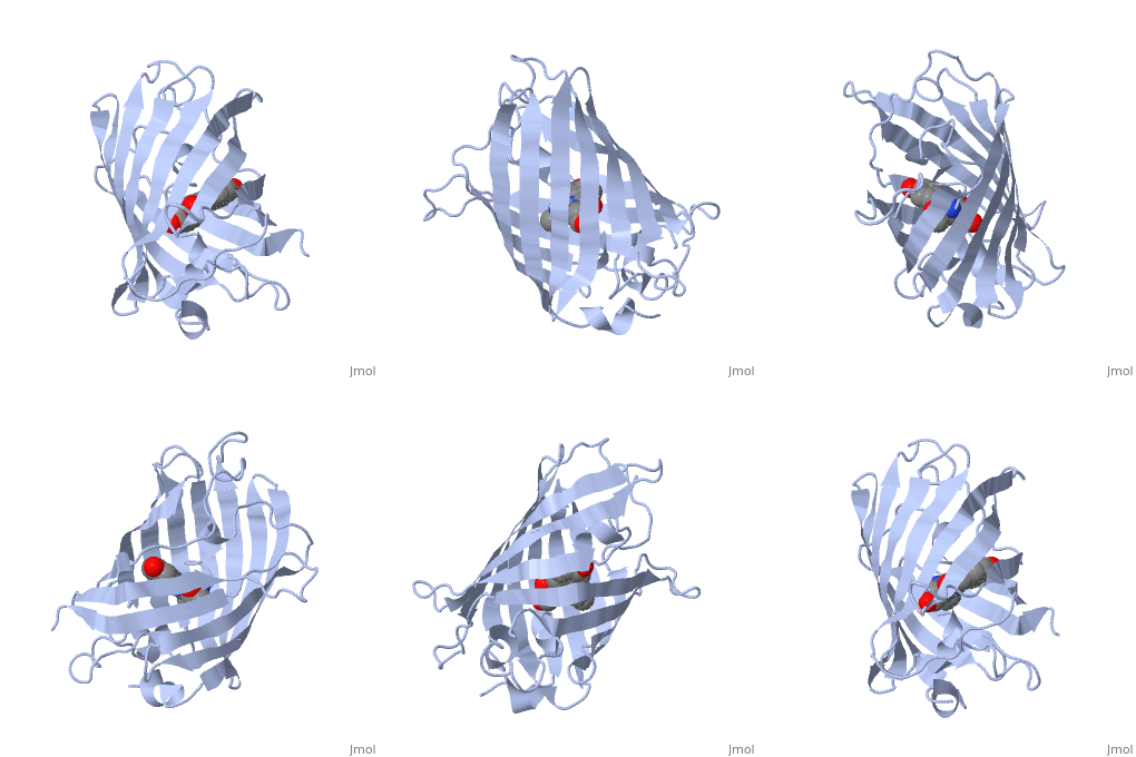
===GREEN FLUORESCENT PROTEIN FROM AEQUOREA VICTORIA=== | ===GREEN FLUORESCENT PROTEIN FROM AEQUOREA VICTORIA=== | ||
| - | |||
{{ABSTRACT_PUBMED_8703075}} | {{ABSTRACT_PUBMED_8703075}} | ||
Revision as of 11:08, 11 March 2013
| |||||||||
| 1ema, resolution 1.90Å () | |||||||||
|---|---|---|---|---|---|---|---|---|---|
| Non-Standard Residues: | , | ||||||||
| |||||||||
| |||||||||
| Resources: | FirstGlance, OCA, RCSB, PDBsum | ||||||||
| Coordinates: | save as pdb, mmCIF, xml | ||||||||
Contents |
GREEN FLUORESCENT PROTEIN FROM AEQUOREA VICTORIA
Template:ABSTRACT PUBMED 8703075
About this Structure
1ema is a 1 chain structure with sequence from Aequorea victoria. The June 2003 RCSB PDB Molecule of the Month feature on Green Fluorescent Protein (GFP) by David S. Goodsell is 10.2210/rcsb_pdb/mom_2003_6. Full crystallographic information is available from OCA.
See Also
- Alyssa Marsico/Sandbox 1
- Devon McCarthy/Sandbox 1
- Gfp vc2
- Green Fluorescent Protein
- Green Fluorescent Protein: Research Tool
- Sandbox104
- User:Joanne Lau/Sandbox 5
- User:Lynmarie K Thompson/2009Chem490
- User:Student
Reference
- Ormo M, Cubitt AB, Kallio K, Gross LA, Tsien RY, Remington SJ. Crystal structure of the Aequorea victoria green fluorescent protein. Science. 1996 Sep 6;273(5280):1392-5. PMID:8703075


