We apologize for Proteopedia being slow to respond. For the past two years, a new implementation of Proteopedia has been being built. Soon, it will replace this 18-year old system. All existing content will be moved to the new system at a date that will be announced here.
3e0u
From Proteopedia
(Difference between revisions)
| Line 1: | Line 1: | ||
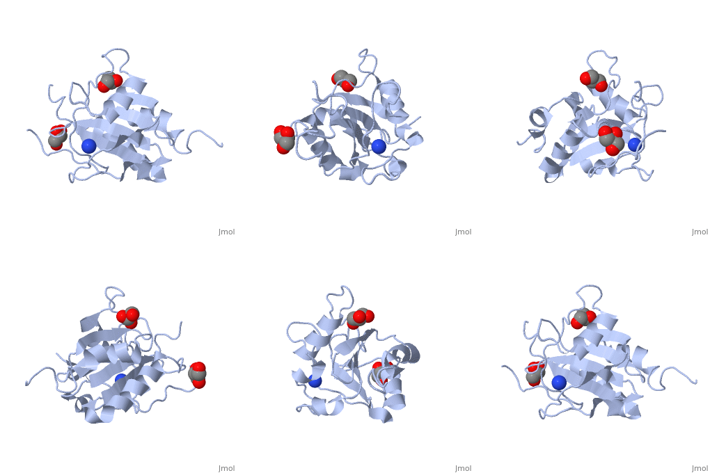
| - | [[ | + | ==Crystal structure of T. cruzi GPX1== |
| + | <StructureSection load='3e0u' size='340' side='right' caption='[[3e0u]], [[Resolution|resolution]] 2.30Å' scene=''> | ||
| + | == Structural highlights == | ||
| + | <table><tr><td colspan='2'>[[3e0u]] is a 1 chain structure with sequence from [http://en.wikipedia.org/wiki/Trypanosoma_cruzi Trypanosoma cruzi]. Full crystallographic information is available from [http://oca.weizmann.ac.il/oca-bin/ocashort?id=3E0U OCA]. For a <b>guided tour on the structure components</b> use [http://oca.weizmann.ac.il/oca-docs/fgij/fg.htm?mol=3E0U FirstGlance]. <br> | ||
| + | </td></tr><tr><td class="sblockLbl"><b>[[Ligand|Ligands:]]</b></td><td class="sblockDat"><scene name='pdbligand=GOL:GLYCEROL'>GOL</scene>, <scene name='pdbligand=NH4:AMMONIUM+ION'>NH4</scene><br> | ||
| + | <tr><td class="sblockLbl"><b>[[Gene|Gene:]]</b></td><td class="sblockDat">GPXI ([http://www.ncbi.nlm.nih.gov/Taxonomy/Browser/wwwtax.cgi?mode=Info&srchmode=5&id=5693 Trypanosoma cruzi])</td></tr> | ||
| + | <tr><td class="sblockLbl"><b>Activity:</b></td><td class="sblockDat"><span class='plainlinks'>[http://en.wikipedia.org/wiki/Glutathione_peroxidase Glutathione peroxidase], with EC number [http://www.brenda-enzymes.info/php/result_flat.php4?ecno=1.11.1.9 1.11.1.9] </span></td></tr> | ||
| + | <tr><td class="sblockLbl"><b>Resources:</b></td><td class="sblockDat"><span class='plainlinks'>[http://oca.weizmann.ac.il/oca-docs/fgij/fg.htm?mol=3e0u FirstGlance], [http://oca.weizmann.ac.il/oca-bin/ocaids?id=3e0u OCA], [http://www.rcsb.org/pdb/explore.do?structureId=3e0u RCSB], [http://www.ebi.ac.uk/pdbsum/3e0u PDBsum]</span></td></tr> | ||
| + | <table> | ||
| + | == Evolutionary Conservation == | ||
| + | [[Image:Consurf_key_small.gif|200px|right]] | ||
| + | Check<jmol> | ||
| + | <jmolCheckbox> | ||
| + | <scriptWhenChecked>select protein; define ~consurf_to_do selected; consurf_initial_scene = true; script "/wiki/ConSurf/e0/3e0u_consurf.spt"</scriptWhenChecked> | ||
| + | <scriptWhenUnchecked>script /wiki/extensions/Proteopedia/spt/initialview01.spt</scriptWhenUnchecked> | ||
| + | <text>to colour the structure by Evolutionary Conservation</text> | ||
| + | </jmolCheckbox> | ||
| + | </jmol>, as determined by [http://consurfdb.tau.ac.il/ ConSurfDB]. You may read the [[Conservation%2C_Evolutionary|explanation]] of the method and the full data available from [http://bental.tau.ac.il/new_ConSurfDB/chain_selection.php?pdb_ID=2ata ConSurf]. | ||
| + | <div style="clear:both"></div> | ||
| + | <div style="background-color:#fffaf0;"> | ||
| + | == Publication Abstract from PubMed == | ||
| + | Current drug therapies against Trypanosoma cruzi, the causative agent of Chagas disease, have limited effectiveness and are highly toxic. T. cruzi-specific metabolic pathways that utilize trypanothione for the reduction of peroxides are being explored as potential novel therapeutic targets. In the present study we solved the X-ray crystal structure of one of the T. cruzi enzymes involved in peroxide reduction, the glutathione peroxidase-like enzyme TcGPXI (T. cruzi glutathione peroxidase-like enzyme I). We also characterized the wild-type, C48G and C96G variants of TcGPXI by NMR spectroscopy and biochemical assays. Our results show that residues Cys48 and Cys96 are required for catalytic activity. In solution, the TcGPXI molecule readily forms a Cys48-Cys96 disulfide bridge and the polypeptide segment containing Cys96 lacks regular secondary structure. NMR spectra of the reduced TcGPXI are indicative of a protein that undergoes widespread conformational exchange on an intermediate time scale. Despite the absence of the disulfide bond, the active site mutant proteins acquired an oxidized-like conformation as judged from their NMR spectra. The protein that was used for crystallization was pre-oxidized by t-butyl hydroperoxide; however, the electron density maps clearly showed that the active site cysteine residues are in the reduced thiol form, indicative of X-ray-induced reduction. Our crystallographic and solution studies suggest a level of structural plasticity in TcGPXI consistent with the requirement of the atypical two-cysteine (2-Cys) peroxiredoxin-like mechanism implied by the behaviour of the Cys48 and Cys96 mutant proteins. | ||
| - | + | Structural insights into the catalytic mechanism of Trypanosoma cruzi GPXI (glutathione peroxidase-like enzyme I).,Patel S, Hussain S, Harris R, Sardiwal S, Kelly JM, Wilkinson SR, Driscoll PC, Djordjevic S Biochem J. 2010 Jan 15;425(3):513-22. PMID:19886864<ref>PMID:19886864</ref> | |
| - | + | From MEDLINE®/PubMed®, a database of the U.S. National Library of Medicine.<br> | |
| - | + | </div> | |
| - | + | ||
| - | + | ||
| - | + | ||
| - | + | ||
==See Also== | ==See Also== | ||
*[[Glutathione peroxidase|Glutathione peroxidase]] | *[[Glutathione peroxidase|Glutathione peroxidase]] | ||
| - | + | == References == | |
| - | == | + | <references/> |
| - | < | + | __TOC__ |
| + | </StructureSection> | ||
[[Category: Glutathione peroxidase]] | [[Category: Glutathione peroxidase]] | ||
[[Category: Trypanosoma cruzi]] | [[Category: Trypanosoma cruzi]] | ||
Revision as of 11:09, 29 September 2014
Crystal structure of T. cruzi GPX1
| |||||||||||


