We apologize for Proteopedia being slow to respond. For the past two years, a new implementation of Proteopedia has been being built. Soon, it will replace this 18-year old system. All existing content will be moved to the new system at a date that will be announced here.
7rsa
From Proteopedia
(Difference between revisions)
| Line 1: | Line 1: | ||
| - | [[Image:7rsa.png|left|200px]] | ||
| - | |||
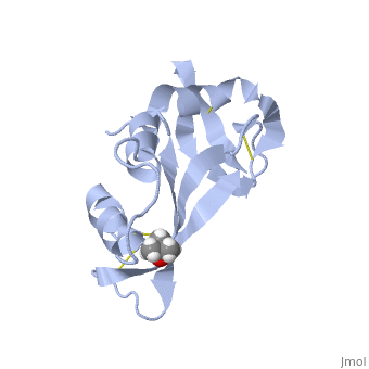
{{STRUCTURE_7rsa| PDB=7rsa | SCENE= }} | {{STRUCTURE_7rsa| PDB=7rsa | SCENE= }} | ||
| - | |||
===STRUCTURE OF PHOSPHATE-FREE RIBONUCLEASE A REFINED AT 1.26 ANGSTROMS=== | ===STRUCTURE OF PHOSPHATE-FREE RIBONUCLEASE A REFINED AT 1.26 ANGSTROMS=== | ||
| - | |||
{{ABSTRACT_PUBMED_3401445}} | {{ABSTRACT_PUBMED_3401445}} | ||
Revision as of 11:08, 11 March 2013
| |||||||||
| 7rsa, resolution 1.26Å () | |||||||||
|---|---|---|---|---|---|---|---|---|---|
| Ligands: | , | ||||||||
| Activity: | Pancreatic ribonuclease, with EC number 3.1.27.5 | ||||||||
| |||||||||
| |||||||||
| |||||||||
| Resources: | FirstGlance, OCA, RCSB, PDBsum | ||||||||
| Coordinates: | save as pdb, mmCIF, xml | ||||||||

Contents |
STRUCTURE OF PHOSPHATE-FREE RIBONUCLEASE A REFINED AT 1.26 ANGSTROMS
Template:ABSTRACT PUBMED 3401445
About this Structure
7rsa is a 1 chain structure with sequence from Bos taurus. Full crystallographic information is available from OCA.
See Also
Reference
- Wlodawer A, Svensson LA, Sjolin L, Gilliland GL. Structure of phosphate-free ribonuclease A refined at 1.26 A. Biochemistry. 1988 Apr 19;27(8):2705-17. PMID:3401445
- Kobe B, Deisenhofer J. Mechanism of ribonuclease inhibition by ribonuclease inhibitor protein based on the crystal structure of its complex with ribonuclease A. J Mol Biol. 1996 Dec 20;264(5):1028-43. PMID:9000628 doi:http://dx.doi.org/10.1006/jmbi.1996.0694
- Richardson JS, Richardson DC. Natural beta-sheet proteins use negative design to avoid edge-to-edge aggregation. Proc Natl Acad Sci U S A. 2002 Mar 5;99(5):2754-9. PMID:11880627 doi:10.1073/pnas.052706099
