Caspase-3 Regulatory Mechanisms
From Proteopedia
(→Caspase-3 Regulation) |
|||
| Line 49: | Line 49: | ||
=== Exosite and Allosteric Site=== | === Exosite and Allosteric Site=== | ||
| - | Since all caspases have very similar active site chemistry, other regions in the protein that can confer either activation or inhibition to the enzyme needs to be explored. For example, exosites in caspase-7 have been identified and | + | Since all caspases have very similar active site chemistry, other regions in the protein that can confer either activation or inhibition to the enzyme needs to be explored. For example, exosites in caspase-7 have been identified and were observed to improve activity (Boucher, Blais et al. 2012). Caspase-7 also has an inhibitory allosteric site that could bind with the small molecule FICA, resulting in a zymogen-like conformation (Hardy, Lam et al. 2004) and abolishing activity. |
| - | Although there are still no evident exosites found in caspase-3, some allosteric sites, most of which are located on the dimer interface, have been interrogated by mutagenesis and | + | Although there are still no evident exosites found in caspase-3, some allosteric sites, most of which are located on the dimer interface, have been interrogated by mutagenesis and have been shown to modulate the activity of caspase-3 or even procaspase-3. Although only procaspase-3 was detected with little activity because the orientation of ILA (prematured L2 loop) and ILB loop cannot form an active site pocket, it is sill quite interesting to discover how these mutations are able to rescue activity of the zymogen form (Bose, Pop et al. 2003). |
| - | One interesting mutation, V266E, improves caspase-3 activity dramatically. Even in the uncleavable procaspase-3 (D5A, D26A, D175A), V266E mutant zymogen is still pseudo-activated, showing a 60-fold increase in activity. Intriguingly, V266E does not undergo | + | One interesting mutation, V266E, improves caspase-3 activity dramatically. Even in the uncleavable procaspase-3 (D5A, D26A, D175A), V266E mutant zymogen is still pseudo-activated, showing a 60-fold increase in activity. Intriguingly, V266E does not undergo many conformational changes around the active site in its cleaved form. Based on the crystal structure of this mutant, the L2’ loop is partially disordered at 185’-180’. Moreover, this active procaspase-3 variant cannot be inhibited by endogenous XIAP like normal cleaved caspase-3. So it provides an option for apoptosis stimuli with intrinsic efficiency. |
It was found recently that changing other critical residues (V266H, Y197C, E124A) on the dimer interface might play an important role on inhibition of caspase-3 by altering hydrogen bond patterns or through a relay of subtle conformational changes that spreads across the whole dimer. | It was found recently that changing other critical residues (V266H, Y197C, E124A) on the dimer interface might play an important role on inhibition of caspase-3 by altering hydrogen bond patterns or through a relay of subtle conformational changes that spreads across the whole dimer. | ||
Revision as of 03:07, 13 December 2012
Contents |
Introduction
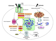
Caspases are cysteine-aspartic acid proteases and are key protein facilitators for the faithful execution of apoptosis or programmed cell death. Dysregulation in the apoptotic pathway has been implicated in a variety of diseases such as neurodegeneration, cancer, heart disease and some metabolic disorders. Because of the crucial role of caspases in the the apoptotic pathway, abnormalities in their functions would cause a haywire in the apoptotic cascade and can be deleterious to the cell. Caspases are thus being considered as therapeutic targets in apoptosis-related diseases.
Any apoptotic signal received by the cell causes the activation of initiator caspases (-8 and -9) by associating with other protein platforms to form a functional holoenzyme. These initiator caspases then cleaves the executioner caspases -3, -6, -7. Caspase-3 specifically functions to cleave both caspase-6 and -7, which in turn cleave their respective targets to induce cell death. Aside from being able to activate caspase-6 and -7, caspase-3 also regulates caspase-9 activity, operating via a feedback loop. This dual action of caspase-3 confers its distinct regulatory mechanisms, resulting a wider extent of its effects in the apoptotic cascade.
Overview of Caspase-3 Structure
| |||||||||||
Caspase-3 Loop Bundle and Active Site
| |||||||||||
Caspase-3 Regulation
| |||||||||||
Proteopedia Page Contributors and Editors (what is this?)
Scott Eron, Banyuhay P. Serrano, Alexander Berchansky, Yunlong Zhao, Jaime Prilusky, Michal Harel


