This old version of Proteopedia is provided for student assignments while the new version is undergoing repairs. Content and edits done in this old version of Proteopedia after March 1, 2026 will eventually be lost when it is retired in about June of 2026.
Apply for new accounts at the new Proteopedia. Your logins will work in both the old and new versions.
2d1r
From Proteopedia
(Difference between revisions)
| Line 1: | Line 1: | ||
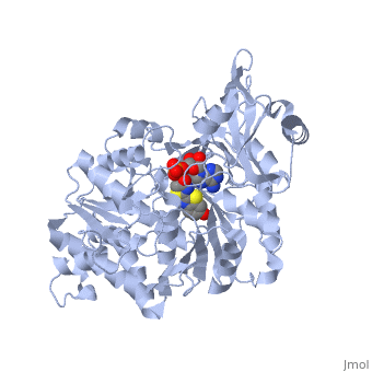
| - | + | ==Crystal structure of the thermostable Japanese firefly Luciferase complexed with OXYLUCIFERIN and AMP== | |
| - | + | <StructureSection load='2d1r' size='340' side='right' caption='[[2d1r]], [[Resolution|resolution]] 1.60Å' scene=''> | |
| - | + | == Structural highlights == | |
| + | <table><tr><td colspan='2'>[[2d1r]] is a 1 chain structure with sequence from [http://en.wikipedia.org/wiki/Luciola_cruciata Luciola cruciata]. Full crystallographic information is available from [http://oca.weizmann.ac.il/oca-bin/ocashort?id=2D1R OCA]. For a <b>guided tour on the structure components</b> use [http://oca.weizmann.ac.il/oca-docs/fgij/fg.htm?mol=2D1R FirstGlance]. <br> | ||
| + | </td></tr><tr><td class="sblockLbl"><b>[[Ligand|Ligands:]]</b></td><td class="sblockDat"><scene name='pdbligand=AMP:ADENOSINE+MONOPHOSPHATE'>AMP</scene>, <scene name='pdbligand=OLU:2-(6-HYDROXY-1,3-BENZOTHIAZOL-2-YL)-1,3-THIAZOL-4(5H)-ONE'>OLU</scene><br> | ||
| + | <tr><td class="sblockLbl"><b>[[Non-Standard_Residue|NonStd Res:]]</b></td><td class="sblockDat"><scene name='pdbligand=CSO:S-HYDROXYCYSTEINE'>CSO</scene></td></tr> | ||
| + | <tr><td class="sblockLbl"><b>[[Related_structure|Related:]]</b></td><td class="sblockDat">[[2d1s|2d1s]], [[2d1t|2d1t]]</td></tr> | ||
| + | <tr><td class="sblockLbl"><b>Activity:</b></td><td class="sblockDat"><span class='plainlinks'>[http://en.wikipedia.org/wiki/Photinus-luciferin_4-monooxygenase_(ATP-hydrolyzing) Photinus-luciferin 4-monooxygenase (ATP-hydrolyzing)], with EC number [http://www.brenda-enzymes.info/php/result_flat.php4?ecno=1.13.12.7 1.13.12.7] </span></td></tr> | ||
| + | <tr><td class="sblockLbl"><b>Resources:</b></td><td class="sblockDat"><span class='plainlinks'>[http://oca.weizmann.ac.il/oca-docs/fgij/fg.htm?mol=2d1r FirstGlance], [http://oca.weizmann.ac.il/oca-bin/ocaids?id=2d1r OCA], [http://www.rcsb.org/pdb/explore.do?structureId=2d1r RCSB], [http://www.ebi.ac.uk/pdbsum/2d1r PDBsum], [http://www.topsan.org/Proteins/RSGI/2d1r TOPSAN]</span></td></tr> | ||
| + | <table> | ||
| + | == Evolutionary Conservation == | ||
| + | [[Image:Consurf_key_small.gif|200px|right]] | ||
| + | Check<jmol> | ||
| + | <jmolCheckbox> | ||
| + | <scriptWhenChecked>select protein; define ~consurf_to_do selected; consurf_initial_scene = true; script "/wiki/ConSurf/d1/2d1r_consurf.spt"</scriptWhenChecked> | ||
| + | <scriptWhenUnchecked>script /wiki/extensions/Proteopedia/spt/initialview01.spt</scriptWhenUnchecked> | ||
| + | <text>to colour the structure by Evolutionary Conservation</text> | ||
| + | </jmolCheckbox> | ||
| + | </jmol>, as determined by [http://consurfdb.tau.ac.il/ ConSurfDB]. You may read the [[Conservation%2C_Evolutionary|explanation]] of the method and the full data available from [http://bental.tau.ac.il/new_ConSurfDB/chain_selection.php?pdb_ID=2ata ConSurf]. | ||
| + | <div style="clear:both"></div> | ||
| + | <div style="background-color:#fffaf0;"> | ||
| + | == Publication Abstract from PubMed == | ||
| + | Fireflies communicate with each other by emitting yellow-green to yellow-orange brilliant light. The bioluminescence reaction, which uses luciferin, Mg-ATP and molecular oxygen to yield an electronically excited oxyluciferin species, is carried out by the enzyme luciferase. Visible light is emitted during relaxation of excited oxyluciferin to its ground state. The high quantum yield of the luciferin/luciferase reaction and the change in bioluminescence colour caused by subtle structural differences in luciferase have attracted much research interest. In fact, a single amino acid substitution in luciferase changes the emission colour from yellow-green to red. Although the crystal structure of luciferase from the North American firefly (Photinus pyralis) has been described, the detailed mechanism for the bioluminescence colour change is still unclear. Here we report the crystal structures of wild-type and red mutant (S286N) luciferases from the Japanese Genji-botaru (Luciola cruciata) in complex with a high-energy intermediate analogue, 5'-O-[N-(dehydroluciferyl)-sulfamoyl]adenosine (DLSA). Comparing these structures to those of the wild-type luciferase complexed with AMP plus oxyluciferin (products) reveals a significant conformational change in the wild-type enzyme but not in the red mutant. This conformational change involves movement of the hydrophobic side chain of Ile 288 towards the benzothiazole ring of DLSA. Our results indicate that the degree of molecular rigidity of the excited state of oxyluciferin, which is controlled by a transient movement of Ile 288, determines the colour of bioluminescence during the emission reaction. | ||
| - | + | Structural basis for the spectral difference in luciferase bioluminescence.,Nakatsu T, Ichiyama S, Hiratake J, Saldanha A, Kobashi N, Sakata K, Kato H Nature. 2006 Mar 16;440(7082):372-6. PMID:16541080<ref>PMID:16541080</ref> | |
| - | + | ||
| + | From MEDLINE®/PubMed®, a database of the U.S. National Library of Medicine.<br> | ||
| + | </div> | ||
==See Also== | ==See Also== | ||
*[[Luciferase|Luciferase]] | *[[Luciferase|Luciferase]] | ||
*[[Luciola cruciata luciferase|Luciola cruciata luciferase]] | *[[Luciola cruciata luciferase|Luciola cruciata luciferase]] | ||
| - | + | == References == | |
| - | == | + | <references/> |
| - | < | + | __TOC__ |
| + | </StructureSection> | ||
[[Category: Luciola cruciata]] | [[Category: Luciola cruciata]] | ||
[[Category: Hiratake, J.]] | [[Category: Hiratake, J.]] | ||
Revision as of 01:54, 30 September 2014
Crystal structure of the thermostable Japanese firefly Luciferase complexed with OXYLUCIFERIN and AMP
| |||||||||||

Categories: Luciola cruciata | Hiratake, J. | Ichiyama, S. | Kato, H. | Kobashi, N. | Nakatsu, T. | RSGI, RIKEN Structural Genomics/Proteomics Initiative. | Sakata, K. | Saldanha, A. | Alpha+beta | Alpha/beta | Beta barrel | Oxidoreductase | Riken structural genomics/proteomics initiative | Rsgi | Structural genomic
