This old version of Proteopedia is provided for student assignments while the new version is undergoing repairs. Content and edits done in this old version of Proteopedia after March 1, 2026 will eventually be lost when it is retired in about June of 2026.
Apply for new accounts at the new Proteopedia. Your logins will work in both the old and new versions.
Aconitase
From Proteopedia
| Line 1: | Line 1: | ||
| + | <StructureSection load='' size='450' side='right' scene='Aconitase/Cv/1' caption=''> | ||
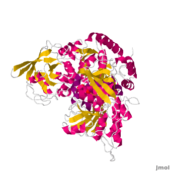
[[Image:1amj.png|left|200px|thumb|Crystal Structure of Bovine Aconitase, [[1amj]]]] | [[Image:1amj.png|left|200px|thumb|Crystal Structure of Bovine Aconitase, [[1amj]]]] | ||
| - | {{STRUCTURE_1amj| PDB=1amj | SIZE=400| SCENE=Aconitase/Cv/1 |right|CAPTION=Bovine aconitase showing the Fe3S4 cluster complex with sulfate, [[1amj]]. α helices (purple) and β strands (yellow). }} | ||
| - | |||
| - | |||
| - | |||
| - | |||
| - | |||
| - | |||
| - | |||
| - | |||
| - | |||
| - | |||
| - | |||
| - | |||
| - | |||
| - | |||
| - | |||
| - | |||
| - | |||
| - | |||
| - | |||
| - | |||
[[Aconitase]] (ACO, EC number [http://www.brenda-enzymes.info/php/result_flat.php4?ecno=4.2.1.3 4.2.1.3]) is an enzymatic domain that confers the ability to catalyse the equilibrium | [[Aconitase]] (ACO, EC number [http://www.brenda-enzymes.info/php/result_flat.php4?ecno=4.2.1.3 4.2.1.3]) is an enzymatic domain that confers the ability to catalyse the equilibrium | ||
:citrate = aconitate + H<sub>2</sub>O = L-isocitrate | :citrate = aconitate + H<sub>2</sub>O = L-isocitrate | ||
| Line 30: | Line 10: | ||
==Structure== | ==Structure== | ||
| - | + | ||
The <scene name='Anthony_Noles_Sandbox/Secondary_structure/1'>secondary structure</scene> consists of numerous alternating alpha helices and beta sheets (SCOP classification α/β alternating). The tertiary structure is somewhat bilobed with the active site in the middle, and, since there is only one subunit, there is no quaternary structure. Aconitase consists of four domains, three of which are tightly packed while the fourth is more flexible. <ref name="Frishman">PMID 8706708</ref> Aconitase contains a <scene name='Anthony_Noles_Sandbox/Fe-scluster/2'>4Fe-4S iron-sulfur cluster</scene>. This iron sulfur cluster does not participate in redox as most do, but holds the OH group of citrate to facilitate its elimination.<ref>PMID:16407072 </ref> It is at this 4Fe-4S site that catalysis occurs and citrate or <scene name='Anthony_Noles_Sandbox/Fe-scluster_bound_isocitrate/8'>isocitrate</scene> is bound. The rest of the <scene name='Anthony_Noles_Sandbox/Fe-scluster_w_active_site/5'>active site (manually rotate this scene to see the proximity of each residue to the 4Fe-4S cluster)</scene> is made up of residues Gln72, Asp100, His101, Asp165, Ser166, His167, His147, Glu262, Asn258, Cys358, Cys421, Cys424, Cys358, Cys421, Asn446, Arg447, Arg452, Asp568, Ser642, Ser643, Arg644, Arg580. <ref name="Beinert">Beinert, H., Kennedy, M. C., Stout, C.D. “Aconitase as Iron−Sulfur Protein, Enzyme, and Iron-Regulatory Protein.” Chem. Rev. 1996, 96, 2335−2373.</ref> | The <scene name='Anthony_Noles_Sandbox/Secondary_structure/1'>secondary structure</scene> consists of numerous alternating alpha helices and beta sheets (SCOP classification α/β alternating). The tertiary structure is somewhat bilobed with the active site in the middle, and, since there is only one subunit, there is no quaternary structure. Aconitase consists of four domains, three of which are tightly packed while the fourth is more flexible. <ref name="Frishman">PMID 8706708</ref> Aconitase contains a <scene name='Anthony_Noles_Sandbox/Fe-scluster/2'>4Fe-4S iron-sulfur cluster</scene>. This iron sulfur cluster does not participate in redox as most do, but holds the OH group of citrate to facilitate its elimination.<ref>PMID:16407072 </ref> It is at this 4Fe-4S site that catalysis occurs and citrate or <scene name='Anthony_Noles_Sandbox/Fe-scluster_bound_isocitrate/8'>isocitrate</scene> is bound. The rest of the <scene name='Anthony_Noles_Sandbox/Fe-scluster_w_active_site/5'>active site (manually rotate this scene to see the proximity of each residue to the 4Fe-4S cluster)</scene> is made up of residues Gln72, Asp100, His101, Asp165, Ser166, His167, His147, Glu262, Asn258, Cys358, Cys421, Cys424, Cys358, Cys421, Asn446, Arg447, Arg452, Asp568, Ser642, Ser643, Arg644, Arg580. <ref name="Beinert">Beinert, H., Kennedy, M. C., Stout, C.D. “Aconitase as Iron−Sulfur Protein, Enzyme, and Iron-Regulatory Protein.” Chem. Rev. 1996, 96, 2335−2373.</ref> | ||
| Line 36: | Line 16: | ||
== Catalytic mechanism of mitochondrial ACO == | == Catalytic mechanism of mitochondrial ACO == | ||
| - | + | Both mAc and cAc are quite similar in their ACO function. Studies, however, concentrated on <scene name='Aconitase/7acn-sf4/1'>the mitochondrial ACO</scene>. ACO is an excellent system for understanding the role of iron-sulfur-clusters in catalysis. The <scene name='Aconitase/7acn-sf4/2'>(4Fe-4S) cofactor is held in place</scene> by three sulfur atoms belonging to the cysteins-385, -448, and -451 <scene name='Aconitase/7acn-morph/3'>which are bound to three of the four</scene> cluster iron atoms. On activation of the enzyme, <scene name='Aconitase/7acn-morph/4'>a fourth iron atom is included in the cluster</scene> together with a water molecule.This Fe4 is free to bind one, two, or three partners, in this reaction always oxygen atoms belonging to other molecules.<ref>PMID:8151704</ref> | |
<!--It is clear that, in order to synthesize L-isocitrate, stereoselective catalysis must occur.--> | <!--It is clear that, in order to synthesize L-isocitrate, stereoselective catalysis must occur.--> | ||
Substrate-free aconitase contains a [4Fe-4S]<sup>2+</sup> cluster with hydroxyl bound to one of the Fe. Upon binding of substrate the bound hydroxyl is protonated. A hydrogen bond from <scene name='Anthony_Noles_Sandbox/His101/3'>His101</scene> to the isocitrate hydroxyl is donated to form water. Alternatively, the proton could be donated by <scene name='Anthony_Noles_Sandbox/His167/3'>His167</scene> as this histidine is hydrogen bonded to a H<sub>2</sub>O molecule. His167 is also hydrogen bonded to the bound H<sub>2</sub>O in the [4Fe-4S] cluster. Both <scene name='Anthony_Noles_Sandbox/His_101_and_167/4'>His101 and His167</scene> are paired with carboxylates (<scene name='Anthony_Noles_Sandbox/Asp100_and_glu262/3'>Asp100 and Glu262</scene>, respectively) and are likely to be protonated. The conformational change associated with substrate binding reorients the cluster. <ref name="Beinert" /> The residue which removes a proton from citrate or isocitrate is <scene name='Anthony_Noles_Sandbox/Ser642/4'>Ser642</scene>. <ref name="Beinert" /> This causes the cis-Aconitate intermediate (seen below), which consists of a double bond, which is a direct result of the deprotonation. Then, there is a rehydration of the double bond of cis-aconitate to form isocitrate (if the original substrate was citrate). To better understand this, consider this process as stages, seen below. | Substrate-free aconitase contains a [4Fe-4S]<sup>2+</sup> cluster with hydroxyl bound to one of the Fe. Upon binding of substrate the bound hydroxyl is protonated. A hydrogen bond from <scene name='Anthony_Noles_Sandbox/His101/3'>His101</scene> to the isocitrate hydroxyl is donated to form water. Alternatively, the proton could be donated by <scene name='Anthony_Noles_Sandbox/His167/3'>His167</scene> as this histidine is hydrogen bonded to a H<sub>2</sub>O molecule. His167 is also hydrogen bonded to the bound H<sub>2</sub>O in the [4Fe-4S] cluster. Both <scene name='Anthony_Noles_Sandbox/His_101_and_167/4'>His101 and His167</scene> are paired with carboxylates (<scene name='Anthony_Noles_Sandbox/Asp100_and_glu262/3'>Asp100 and Glu262</scene>, respectively) and are likely to be protonated. The conformational change associated with substrate binding reorients the cluster. <ref name="Beinert" /> The residue which removes a proton from citrate or isocitrate is <scene name='Anthony_Noles_Sandbox/Ser642/4'>Ser642</scene>. <ref name="Beinert" /> This causes the cis-Aconitate intermediate (seen below), which consists of a double bond, which is a direct result of the deprotonation. Then, there is a rehydration of the double bond of cis-aconitate to form isocitrate (if the original substrate was citrate). To better understand this, consider this process as stages, seen below. | ||
| Line 57: | Line 37: | ||
The Citric Acid Cycle works in such a way that the product of one reaction becomes the reactant of another, with different enzymes catalyzing each reaction. Aconitase is one such enzyme. Some of these enzymes are tightly regulated, either activated or inhibited, by the concentration of reactant, product, ATP or NADH, and thus are rate-determining. Aconitase is not one of the three rate-determining enzymes of the Citric Acid Cycle as its ΔG is not negative (ΔG°′≈5 kJ/mol and ΔG≈0 kJ/mol).<ref name="Voet" /> Aconitase functions close to equilibrium and the rate of citrate consumption depends on the activity of NAD<sup>+</sup>-dependent isocitrate dehydrogenase, which is one of the three rate-determining enyzmes. Isocitrate dehydrogenase uses the product of the reaction aconitase catalyzes. Both Citrate synthase and Isocitrate dehydogenase are inhibited by NADH concentration, but aconitase itself is not.<ref name="Voet" /> Since the rate of aconitase depends on the activity of NAD<sup>+</sup>-dependent isocitrate dehydrogenase, then citrate could build up on the reactant side, which would then inhibit the enzyme of the previous step, citrate synthase. An illustration of this is seen below, with the boxes representing the enzymes that are catalyzing each reaction. This is a common example of how the Citric Acid Cycle works in order to produce ATP without wasting resources. Similar inhibition/activation of enzymes occurs based on concentrations of ATP, NADH, Calcium, CoA, and others. | The Citric Acid Cycle works in such a way that the product of one reaction becomes the reactant of another, with different enzymes catalyzing each reaction. Aconitase is one such enzyme. Some of these enzymes are tightly regulated, either activated or inhibited, by the concentration of reactant, product, ATP or NADH, and thus are rate-determining. Aconitase is not one of the three rate-determining enzymes of the Citric Acid Cycle as its ΔG is not negative (ΔG°′≈5 kJ/mol and ΔG≈0 kJ/mol).<ref name="Voet" /> Aconitase functions close to equilibrium and the rate of citrate consumption depends on the activity of NAD<sup>+</sup>-dependent isocitrate dehydrogenase, which is one of the three rate-determining enyzmes. Isocitrate dehydrogenase uses the product of the reaction aconitase catalyzes. Both Citrate synthase and Isocitrate dehydogenase are inhibited by NADH concentration, but aconitase itself is not.<ref name="Voet" /> Since the rate of aconitase depends on the activity of NAD<sup>+</sup>-dependent isocitrate dehydrogenase, then citrate could build up on the reactant side, which would then inhibit the enzyme of the previous step, citrate synthase. An illustration of this is seen below, with the boxes representing the enzymes that are catalyzing each reaction. This is a common example of how the Citric Acid Cycle works in order to produce ATP without wasting resources. Similar inhibition/activation of enzymes occurs based on concentrations of ATP, NADH, Calcium, CoA, and others. | ||
[[ Image:Regulation.JPG]] | [[ Image:Regulation.JPG]] | ||
| - | |||
| - | |||
== Cytosolic aconitase and its other function == | == Cytosolic aconitase and its other function == | ||
<applet load='Morph_2ipy-2b3x.pdb.gz' scene='Aconitase/2ipy-total/2' size='400' frame='true' align='right' caption="Cytosolic aconitase from rabbit (bound to RNA) and human (with Fe4S4 cluster), from PDB [[2ipy]] and [[2b3x]]." />A specialty of cAc is that in mammals it has developed a <scene name='Aconitase/2ipy-total/2'>second function</scene> as inhibitor of <scene name='Aconitase/2ipy-rna/1'>those mRNA</scene> that carry an <scene name='Aconitase/2ipy-rna-ire/1'>iron-responsive element (IRE)</scene>. Therefore, the cytosolic cAc is named IREBP for IRE-binding protein when this function is talked about. Only one of the two functions is active, depending on whether <scene name='Aconitase/2b3x-cluster/1'>the (4Fe-4S) cofactor</scene> is present in the molecule: it's essential for <scene name='Aconitase/2b3x-total/1'>the ACO function</scene>. You can see, by <scene name='Aconitase/Morph/2'>looking at the morph</scene>, how much the enzyme structure differs between those two functions. | <applet load='Morph_2ipy-2b3x.pdb.gz' scene='Aconitase/2ipy-total/2' size='400' frame='true' align='right' caption="Cytosolic aconitase from rabbit (bound to RNA) and human (with Fe4S4 cluster), from PDB [[2ipy]] and [[2b3x]]." />A specialty of cAc is that in mammals it has developed a <scene name='Aconitase/2ipy-total/2'>second function</scene> as inhibitor of <scene name='Aconitase/2ipy-rna/1'>those mRNA</scene> that carry an <scene name='Aconitase/2ipy-rna-ire/1'>iron-responsive element (IRE)</scene>. Therefore, the cytosolic cAc is named IREBP for IRE-binding protein when this function is talked about. Only one of the two functions is active, depending on whether <scene name='Aconitase/2b3x-cluster/1'>the (4Fe-4S) cofactor</scene> is present in the molecule: it's essential for <scene name='Aconitase/2b3x-total/1'>the ACO function</scene>. You can see, by <scene name='Aconitase/Morph/2'>looking at the morph</scene>, how much the enzyme structure differs between those two functions. | ||
| - | |||
Along with serving as a catalyst, aconitase is a member of the iron regulatory protien-1 (IRP-1) family. These enzymes have been found to play a role in regulatory RNA-binding proteins. This suggests a novel role for Fe-S clusters as post-translational regulatory switches.<ref name="Frishman" /> | Along with serving as a catalyst, aconitase is a member of the iron regulatory protien-1 (IRP-1) family. These enzymes have been found to play a role in regulatory RNA-binding proteins. This suggests a novel role for Fe-S clusters as post-translational regulatory switches.<ref name="Frishman" /> | ||
| - | + | ||
| + | |||
== 3D structures of Aconitase== | == 3D structures of Aconitase== | ||
| + | </StructureSection> | ||
| + | __NOTOC__ | ||
Updated on {{REVISIONDAY2}}-{{MONTHNAME|{{REVISIONMONTH}}}}-{{REVISIONYEAR}} | Updated on {{REVISIONDAY2}}-{{MONTHNAME|{{REVISIONMONTH}}}}-{{REVISIONYEAR}} | ||
Revision as of 11:25, 16 October 2013
| |||||||||||


Updated on 16-October-2013
ACO
1b0k – pACO (mutant) – pig
5acn – pACO+Fe3S4
6acn - pACO+Fe4S4
1amj, 1nit – cACO - cow
ACO+citrate
1c96 - pACO (mutant)+citrate
1b0m - pACO (mutant)+fluorocitrate
ACO+aconitate
1fgh – cACO+4-hydroxy-aconitate
1aco – cACO+transaconitate
1nis - cACO+transaconitate+nitrocitrate
ACO+isocitrate
7acn - pACO +isocitrate
1c97, 1b0j - pACO (mutant)+isocitrate
1ami, 8acn – cACO+isocitrate
ACO1
2b3x, 2b3y – hACO1 – human
2ipy, 3snp – rACO1 (mutant)+ferritin H IRE-RNA – rabbit
ACO2
1l5j – ACO2 – Escherichia coli
Literature
- M. Claire Kennedy and Helmut Beinert: IX.4. Aconitase. in Ivano Bertini, Harry B. Gray, Edward I. Stiefel, Joan Selverstone Valentine (eds.): Biological Inorganic Chemistry: Structure and Reactivity. University Science Books, Herndon 2006. ISBN 1891389432 pp.209--
Additional Resources
For additional information, see: Carbohydrate Metabolism
References
- ↑ Zheng L, Kennedy MC, Beinert H, Zalkin H. Mutational analysis of active site residues in pig heart aconitase. J Biol Chem. 1992 Apr 15;267(11):7895-903. PMID:1313811
- ↑ 2.0 2.1 Frishman D, Hentze MW. Conservation of aconitase residues revealed by multiple sequence analysis. Implications for structure/function relationships. Eur J Biochem. 1996 Jul 1;239(1):197-200. PMID:8706708
- ↑ Dupuy J, Volbeda A, Carpentier P, Darnault C, Moulis JM, Fontecilla-Camps JC. Crystal structure of human iron regulatory protein 1 as cytosolic aconitase. Structure. 2006 Jan;14(1):129-39. PMID:16407072 doi:10.1016/j.str.2005.09.009
- ↑ 4.0 4.1 4.2 Beinert, H., Kennedy, M. C., Stout, C.D. “Aconitase as Iron−Sulfur Protein, Enzyme, and Iron-Regulatory Protein.” Chem. Rev. 1996, 96, 2335−2373.
- ↑ Lauble H, Kennedy MC, Beinert H, Stout CD. Crystal structures of aconitase with trans-aconitate and nitrocitrate bound. J Mol Biol. 1994 Apr 8;237(4):437-51. PMID:8151704 doi:http://dx.doi.org/10.1006/jmbi.1994.1246
- ↑ 6.0 6.1 6.2 6.3 Voet, Donald, Judith G. Voet, and Charlotte W. Pratt. Fundamentals of Biochemistry Life at the Molecular Level. New York: John Wiley & Sons, 2008. p. 578-579. Print.
- ↑ 7.0 7.1 Flint, DH., and Allen, RM. "Iron-sulfur protein with nonredox functions.” Chem. Rev. 1996, 96, 2315−2334.
External links
Proteopedia Page Contributors and Editors (what is this?)
Michal Harel, Alexander Berchansky, Ralf Stephan, David Canner, Joel L. Sussman, Jaime Prilusky, Anthony Noles, Angel Herraez, Eran Hodis
