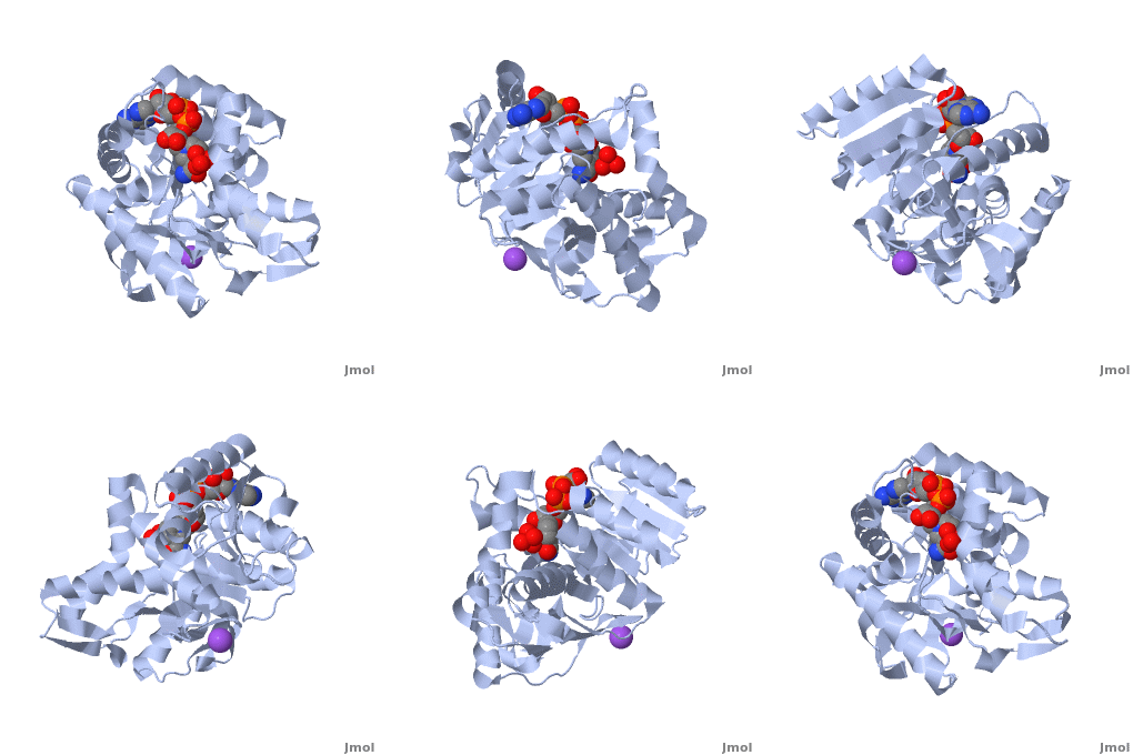
Malate dehydrogenase
From Proteopedia
| Line 1: | Line 1: | ||
| + | <StructureSection load='2x0i' size='450' side='right' scene='Malate_dehydrogenase/Cv/1' caption=''> | ||
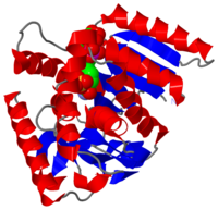
[[Image:2x0i.png|left|200px|thumb|Crystal Structure of Malate Dehydrogenase [[2x0i]]]] | [[Image:2x0i.png|left|200px|thumb|Crystal Structure of Malate Dehydrogenase [[2x0i]]]] | ||
| - | {{STRUCTURE_2j5r| PDB=2j5r | SIZE=400| SCENE= |right|CAPTION=Malate dehydrogenase tetramer complex with Cl- ion [[2j5r]] }} | ||
| - | |||
| Line 23: | Line 22: | ||
{{TOC limit|limit=2}} | {{TOC limit|limit=2}} | ||
{{Clear}} | {{Clear}} | ||
| - | <applet load='2x0i' size='400' frame='true' align='right' scene='Malate_dehydrogenase/Cv/1' / caption= 'Malate dehydrogenase monomer complex with NADH, sulfate and Na+ ion, [[2xi0]]'> | ||
| - | |||
==Structure== | ==Structure== | ||
The secondary structure of a single subunit contains a <scene name='Malate_dehydrogenase/Beta_sheeting_backbone/1'>9 beta sheet parallel backbone</scene> wrapped by <scene name='Malate_dehydrogenase/Alpha_wrapping_betas/1'>9 large alpha helices</scene>. Near the sodium bound end, 4 small anti-parallel beta sheets and 1 small alpha helix enable a turn in the residue chain<scene name='Jake_Ezell_Sandbox_2/Small_turn/1'>(small turn)</scene>. Opposite the sodium bound ligand, 6 alpha helices point towards a common point, three on each side of the beta sheet backbone. The alpha helices form a <scene name='Jake_Ezell_Sandbox_2/Small_groove_nad/1'>small groove</scene> for a NAD+ cofactor to attach near the beta sheeting. The structure most nearly resembles an alternating alpha/beta classification. As for the 3D structure, the enzyme forms a sort of | The secondary structure of a single subunit contains a <scene name='Malate_dehydrogenase/Beta_sheeting_backbone/1'>9 beta sheet parallel backbone</scene> wrapped by <scene name='Malate_dehydrogenase/Alpha_wrapping_betas/1'>9 large alpha helices</scene>. Near the sodium bound end, 4 small anti-parallel beta sheets and 1 small alpha helix enable a turn in the residue chain<scene name='Jake_Ezell_Sandbox_2/Small_turn/1'>(small turn)</scene>. Opposite the sodium bound ligand, 6 alpha helices point towards a common point, three on each side of the beta sheet backbone. The alpha helices form a <scene name='Jake_Ezell_Sandbox_2/Small_groove_nad/1'>small groove</scene> for a NAD+ cofactor to attach near the beta sheeting. The structure most nearly resembles an alternating alpha/beta classification. As for the 3D structure, the enzyme forms a sort of | ||
| Line 34: | Line 31: | ||
==Evolutionary Divergence== | ==Evolutionary Divergence== | ||
The evolutionary past of MDH shows a divergence to form lactate dehydrogenase (LDH) which functions in a very similar way to MDH. Although there is a very low sequence conservation among MDH and LDH’s [http://blast.ncbi.nlm.nih.gov/Blast.cgi] the structure of the enzyme has remained relatively conserved. The key difference between the two is in the substrate: LDH catalyzes pyruvate to lactate. | The evolutionary past of MDH shows a divergence to form lactate dehydrogenase (LDH) which functions in a very similar way to MDH. Although there is a very low sequence conservation among MDH and LDH’s [http://blast.ncbi.nlm.nih.gov/Blast.cgi] the structure of the enzyme has remained relatively conserved. The key difference between the two is in the substrate: LDH catalyzes pyruvate to lactate. | ||
| + | </StructureSection> | ||
| + | __NOTOC__ | ||
== 3D Structures of Malate Dehydrogenase == | == 3D Structures of Malate Dehydrogenase == | ||
Revision as of 13:35, 14 August 2013
| |||||||||||
3D Structures of Malate Dehydrogenase
Updated on 14-August-2013
The holo-MDH contains NAD or its derivatives while the apo-MDH lacks it.
Holo-MDH
2x0r – HmMDH (mutant)+NAD - Haloarcula marismortui
1o6z - HmMDH (mutant)+NADH
1hlp – HmMDH+NAD
1x0i – AfMDH+NADH – Archaeoglobus fulgidus
2x0j - AfMDH+etheno-NAD
1hlp – HmMDH+NAD
1x0i – AfMDH+NADH
2x0j - AfMDH+etheno-NAD
1ib6, 1ie3 – EcMDH (mutant)+NAD - Escherichia coli
1emd – EcMDH+NAD+citrate
3i0p – MDH+NAD – Entamoeba histolytica
3gvh – BmMDH+NAD – Brucella melitensis
3gvi - BmMDH+ADP
2hjr – MDH+adenosine diphosphoribose – Cryptosporidium parvum
2dfd – MDH+NAD – human type 2
1wze – TfMDH (mutant)+NAD – Thermus flavus
1wzi - TfMDH (mutant)+NDP
1bdm - TfMDH (mutant)+beta-6-hydroxy-1,4,5,6-tetrahydronicotinamide adenine dinucleotide
1bmd – TfMDH+NAD
1y7t – TtMDH+NADPH – Thermus thermophilus
2cvq - TtMDH+NADP
1v9n – MDH+NADPH – Pyrococcus horikoshii
1z2i – MDH+NAD – Agrobacterium tumefaciens
1uxg, 1uxh, 1uxi, 1uxj, 1uxk, 1ur5 – MDH (mutant)+NAD – Chloroflexus aurantiacus
1guz, 1guy, 1gv0 – CvMDH+NAD – Chlorobium vibrioforme
1civ – MDH+NADP – Flaveria bidentis
1b8u, 1b8v – AaMDH+NAD - Aquaspirillum arcticum
5mdh – SsMDH+NAD+alpha-ketomalonic acid – Sus scrofa
4mdh – SsMDH+NAD
4i1i – LmMd + NAD – Leishmania major
apo-MDH
2j5r, 2j5k, 2j5q, 1d3a – HmMDH
2hlp – HmMDH (mutant)
3hhp, 2pwz – EcMDH
3fi9 – MDH – Porphyromonas gingivalis
3d5t - MDH – Burkholderia pseudomallei
2d4a – MDH – Aeropyrum pernix
1iz9 - TtMDH
1sev, 1smk – MDH – Citrullus lanatus
1gv1 – CvMDH
1b8p – AaMDH
7mdh – MDH – Sorgum bicolor
1mld – SsMDH
2cmd - EcMd+citrate
3nep – Md – Salinibacter ruber
3p7m – Md – Francisella tularensis
3tl2 – Md – Bacillus anthracis
4e0b – Md – Vibrio vulnificus
4h7p - LmMd
Additional Resources
For additional information, see: Carbohydrate Metabolism
References
- ↑ Minarik P, Tomaskova N, Kollarova M, Antalik M. Malate dehydrogenases--structure and function. Gen Physiol Biophys. 2002 Sep;21(3):257-65. PMID:12537350
- ↑ Matsuda T, Takahashi-Yanaga F, Yoshihara T, Maenaka K, Watanabe Y, Miwa Y, Morimoto S, Kubohara Y, Hirata M, Sasaguri T. Dictyostelium Differentiation-Inducing Factor-1 Binds to Mitochondrial Malate Dehydrogenase and Inhibits Its Activity. J Pharmacol Sci. 2010 Feb 20. PMID:20173310
- ↑ Matsuda T, Takahashi-Yanaga F, Yoshihara T, Maenaka K, Watanabe Y, Miwa Y, Morimoto S, Kubohara Y, Hirata M, Sasaguri T. Dictyostelium Differentiation-Inducing Factor-1 Binds to Mitochondrial Malate Dehydrogenase and Inhibits Its Activity. J Pharmacol Sci. 2010 Feb 20. PMID:20173310
- ↑ Musrati RA, Kollarova M, Mernik N, Mikulasova D. Malate dehydrogenase: distribution, function and properties. Gen Physiol Biophys. 1998 Sep;17(3):193-210. PMID:9834842
- ↑ Boernke WE, Millard CS, Stevens PW, Kakar SN, Stevens FJ, Donnelly MI. Stringency of substrate specificity of Escherichia coli malate dehydrogenase. Arch Biochem Biophys. 1995 Sep 10;322(1):43-52. PMID:7574693 doi:http://dx.doi.org/10.1006/abbi.1995.1434
- ↑ Plancarte A, Nava G, Mendoza-Hernandez G. Purification, properties, and kinetic studies of cytoplasmic malate dehydrogenase from Taenia solium cysticerci. Parasitol Res. 2009 Jul;105(1):175-83. Epub 2009 Mar 10. PMID:19277715 doi:10.1007/s00436-009-1380-6
- ↑ Goward CR, Nicholls DJ. Malate dehydrogenase: a model for structure, evolution, and catalysis. Protein Sci. 1994 Oct;3(10):1883-8. PMID:7849603 doi:http://dx.doi.org/10.1002/pro.5560031027
Proteopedia Page Contributors and Editors (what is this?)
Michal Harel, Alexander Berchansky, Jake Ezell, Joel L. Sussman, Joshua Johnson, Angel Herraez, Jaime Prilusky, David Canner


