P53
From Proteopedia
| Line 1: | Line 1: | ||
| + | <StructureSection load='1tup' size='350' side='right' scene='' caption=''> | ||
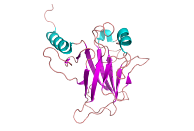
[[Image:P53_DNAbd.png | 400 px | thumb ]] | [[Image:P53_DNAbd.png | 400 px | thumb ]] | ||
| + | {{Clear}} | ||
'''p53 Tumor Suppressor Protein''' | '''p53 Tumor Suppressor Protein''' | ||
__NOTOC__ | __NOTOC__ | ||
| Line 36: | Line 38: | ||
---- | ---- | ||
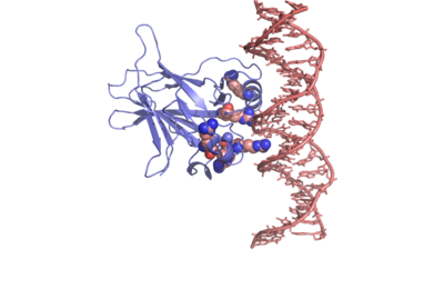
[[Image:P53_DNA.png | 400 px | thumb]] | [[Image:P53_DNA.png | 400 px | thumb]] | ||
| + | {{Clear}} | ||
For more details see [[P53-DNA Recognition]]. | For more details see [[P53-DNA Recognition]]. | ||
| Line 49: | Line 52: | ||
---- | ---- | ||
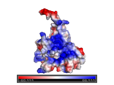
[[Image:P53_surface_charge.png | left | 400 px | thumb]] | [[Image:P53_surface_charge.png | left | 400 px | thumb]] | ||
| - | + | {{Clear}} | |
| Line 63: | Line 66: | ||
| - | + | ||
<scene name='Sandbox/P53_dna_binding_domain/1'>Click Here to view a Three-dimensional Representation of the DNA-binding Domain Bound to DNA</scene> | <scene name='Sandbox/P53_dna_binding_domain/1'>Click Here to view a Three-dimensional Representation of the DNA-binding Domain Bound to DNA</scene> | ||
| Line 72: | Line 75: | ||
zinc plays a role in stabilizing the two loops through | zinc plays a role in stabilizing the two loops through | ||
coordination. The Zn has been represented as a red sphere in the figure at the right. | coordination. The Zn has been represented as a red sphere in the figure at the right. | ||
| + | </StructureSection> | ||
| + | __NOTOC__ | ||
==3D structures of p53== | ==3D structures of p53== | ||
Revision as of 14:23, 23 December 2013
| |||||||||||
3D structures of p53
Updated on 23-December-2013
p53 DNA-binding domain
2xwr, 2ocj, 2ybg – h-p53 DBD – human
2fej - h-p53 DBD - NMR
2wgx, 3d05, 3d06, 3d07, 3d08, 3d09, 2qvq, 2qxa, 2qxb, 2qxc, 2pcx, 2j1w, 2j1x, 2j1y, 2j1z, 2j20, 2j21, 2bim, 2bin, 2bio, 2bip, 2biq, 1uol, 4kvp, 4lo9, 4loe, 4lof - h-p53 DBD (mutant)
1hs5 - h-p53 residues 324-357 - NMR
3q01 - h-p53 DBD + TD
2ioi, 2ioo, 1hu8 – m-p53 DBD – mouse
1t4w – p53-like DBD – Caenoharbditis elegans
p53 DNA-binding domain complex with DNA
3igk, 3igl, 3kz8, 3kmd, 2ac0, 2ady, 2ahi, 2ata, 1tsr, 1tup, 4hje – h-p53 DBD + DNA
3d0a, 3ts8 - h-p53 DBD (mutant) + DNA
3q05, 3q06 - h-p53 DBD + TD + DNA
3exj, 3exl, 2p52, 2geq – m-p53 DBD + DNA
p53 DNA-binding domain complex with small molecule
2x0u, 2x0v, 2x0w, 4agl, 4agm, 4agn, 4ago, 4agp, 4agq – h-p53 DBD (mutant) + benzene derivative
3zme - h-p53 DBD (mutant) + pyrazol derivative
2vuk - h-p53 DBD + drug
2iom - m-p53 DBD + propanol
p53 DNA-binding domain complex with protein
2k8f – h-p53 residues 1-39 + histone acetyltransferase (mutant) – NMR
2h1l - h-p53 DBD + large T antigen
1ycs - h-p53 DBD + 53BP2
1gzh, 1kzy - h-p53 DBD + tumor suppressor p53-binding protein
p53 transactivation domain
2z5s, 2z5t - h-p53 TAD + MDM4 protein
1ycq, 1ycr - h-p53 TAD + MDM2 protein
2l14 – h-p53 TAD + CREB-binding protein
2gs0 – h-p53 TAD + RNA polymerase II transcription factor
p53 tetramerization domain
2j0z, 3sak, 1sae, 1saf, 1sah, 1saj, 1sak, 1sal, 1olh, 1pes, 1pet, 1olg – h-p53 TD – NMR
1c26, 1aie - h-p53 TD
2j10, 2j11, 1a1u - h-p53 TD (mutant) – NMR
Additional Resources
For additional information, see: Cancer
For additional information, see: Oncogenes
Proteopedia Page Contributors and Editors (what is this?)
Joel L. Sussman, Michal Harel, Eran Hodis, Mary Ball, Alexander Berchansky, David Canner


