C-di-GMP specific phosphodiesterases
From Proteopedia
(Difference between revisions)
| Line 1: | Line 1: | ||
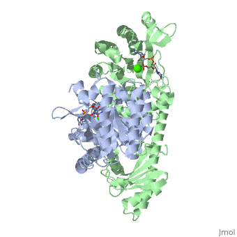
| - | <StructureSection load='2w27' size='450' side='right' scene='' caption='C-di-GMP specific phosphodiesterase complex with guanosine-5 | + | <StructureSection load='2w27' size='450' side='right' scene='' caption='C-di-GMP specific phosphodiesterase complex with guanosine-5-monophosphate and Ca+2 ions (PDB code [[2w27]])'> |
[[C-di-GMP signaling|Back to C-di-GMP signaling]] | [[C-di-GMP signaling|Back to C-di-GMP signaling]] | ||
Revision as of 11:13, 4 December 2013
| |||||||||||
3D structure of YukI
Updated on 04-December-2013
2bas – BsYkuI – Bacillus subtilis
2w27 – BsYkuI + C-di-GMP
3D structure of BlrP1
3gfx, 3gfy, 3gfz, 3gg0, 3gg1 – KpBlrP1 + C-di-GMP - Klebsiella pneumoniae
2kb2 - KpBlrP1 BLUF domain - NMR
3D structures of phosphodiesterase
References
YkuI structure 2w27:
- Minasov G, Padavattan S, Shuvalova L, Brunzelle JS, Miller DJ, Basle A, Massa C, Collart FR, Schirmer T, Anderson WF. Crystal structures of YkuI and its complex with second messenger cyclic Di-GMP suggest catalytic mechanism of phosphodiester bond cleavage by EAL domains. J Biol Chem. 2009 May 8;284(19):13174-84. Epub 2009 Feb 24. PMID:19244251 doi:10.1074/jbc.M808221200
BlrP1 structure 3gg0:
- Barends TR, Hartmann E, Griese JJ, Beitlich T, Kirienko NV, Ryjenkov DA, Reinstein J, Shoeman RL, Gomelsky M, Schlichting I. Structure and mechanism of a bacterial light-regulated cyclic nucleotide phosphodiesterase. Nature. 2009 Jun 18;459(7249):1015-8. PMID:19536266 doi:10.1038/nature07966
Proteopedia Page Contributors and Editors (what is this?)
Michal Harel, Joel L. Sussman, Alexander Berchansky, Tilman Schirmer
